
What Problem Does pixMac Solve?
Macrophage-based in vitro studies have traditionally relied on either primary human monocyte-derived macrophages (HMDMs) or immortalised monocytic cell lines, each with important limitations. HMDMs are generally regarded as the most physiologically relevant option, but they are terminally differentiated, non-proliferative, relatively difficult to genetically manipulate and subject to substantial donor-to-donor variability, which can limit reproducibility and scalability. The THP-1 monocytic leukaemia cell line provides a more consistent and readily expandable alternative, but differs substantially from primary macrophages in both genotype and phenotype. THP-1 cells are karyotypically abnormal and exhibit a relatively immature macrophage phenotype, while differentiation typically requires phorbol 12-myristate 13-acetate (PMA), a strong protein kinase C activator that can itself alter inflammatory signalling and influence downstream immune readouts. Together, these limitations create a need for a scalable, genetically defined and functionally mature human macrophage system with improved experimental reproducibility.
iPSC-derived macrophages (iPSDMs) have emerged as a valuable alternative to primary monocyte-derived macrophages and immortalised monocytic cell lines, providing a genotype-defined, scalable and renewable source of human macrophages derived from a well-characterised starting cell population. This offers greater experimental consistency while retaining the ability to investigate donor-specific or engineered genetic backgrounds. pixMac is pixlbio’s implementation of this approach and has been functionally characterised and benchmarked directly against THP-1-derived macrophages to assess macrophage identity, maturation and immune responsiveness.
Is pixMac Really a Macrophage?
Macrophage identity was established using complementary morphological, transcriptional, protein-level and functional readouts rather than relying on a single marker. Morphologically, pixMac acquire the expected macrophage phenotype, transitioning from smaller, rounded monocyte-like cells to larger, adherent cells with more irregular morphology. At transcriptional level, expression of the macrophage-associated marker CD68 increases during maturation, while CD14 expression decreases, consistent with progression toward a macrophage phenotype. Immunofluorescence analysis further confirms co-expression of the pan-leukocyte marker CD45 and the myeloid/macrophage marker IBA1 across the cell population. Importantly, this molecular characterisation is supported by functional evidence, with pixMac demonstrating efficient phagocytic uptake of fluorescent bead targets, confirming acquisition of a key macrophage-specific function.
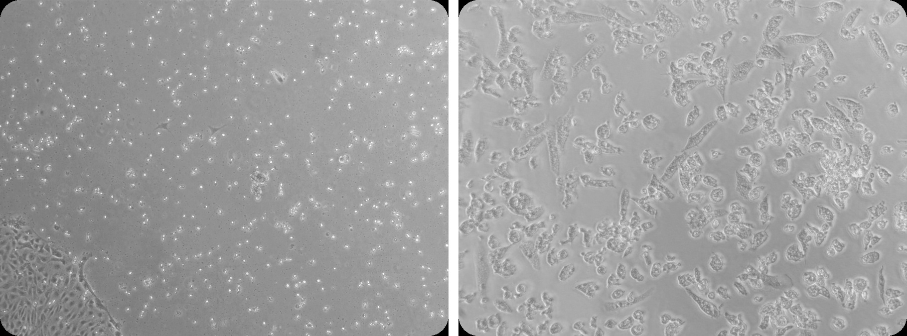
Brightfield morphology: iPSC-derived monocytes (left) transitioning to mature pixMac macrophages (right).
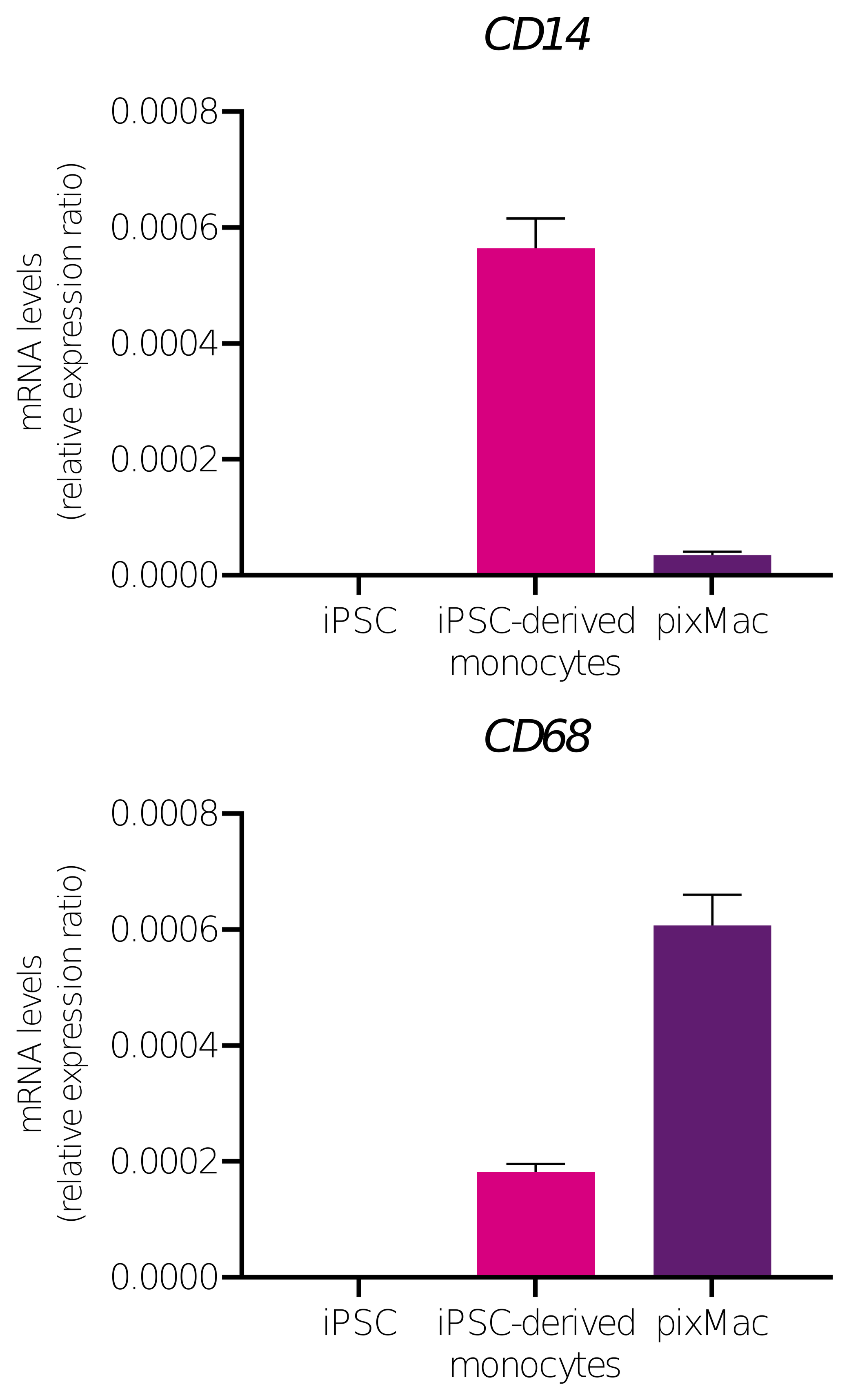
qPCR marker expression: the monocyte marker CD14 declines and the macrophage marker CD68 rises across differentiation from iPSC to iPSC-derived monocytes to pixMac.
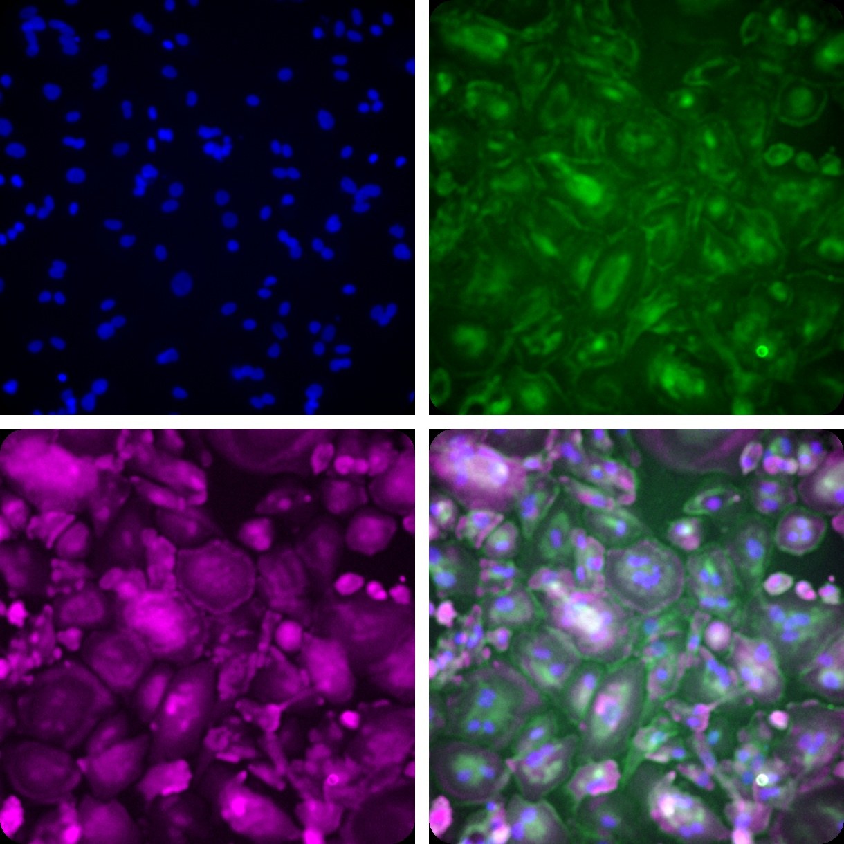
Immunofluorescence: DAPI (nuclei), CD45, IBA1, and overlay. CD45/IBA1 co-expression confirms macrophage identity.
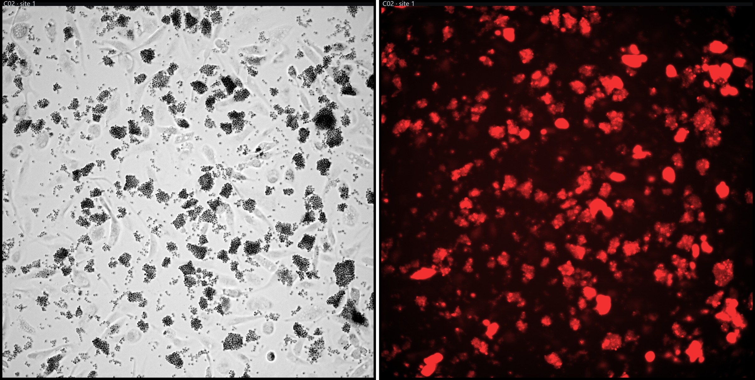
Phagocytosis assay: brightfield (left) and fluorescent red bead uptake (right), confirming functional phagocytic activity.
Can pixMac Polarise?
Macrophage identity alone is insufficient to establish functional maturity; an additional criterion is the ability to respond appropriately to defined activating stimuli. This is a known limitation of commonly used immortalised monocytic cell lines, with comparative studies of THP-1 and U937 cells showing differential biases in their responses to pro- and anti-inflammatory polarisation conditions. pixMac was therefore evaluated for its ability to respond to both activation states, with M0 macrophages exposed for 24 hours to IFNγ and LPS to induce a pro-inflammatory phenotype or to IL-4 to induce an alternatively activated phenotype.
Under pro-inflammatory conditions, pixMac showed increased expression of IL1B and TNF, accompanied by robust secretion of IL-6 and TNF into the culture medium, consistent with a strong inflammatory response. Under IL-4 stimulation, pixMac showed increased IL10 expression at both transcript and protein levels. In contrast, IL10 expression was reduced under pro-inflammatory conditions, demonstrating distinct transcriptional and secretory responses to the two activation states.
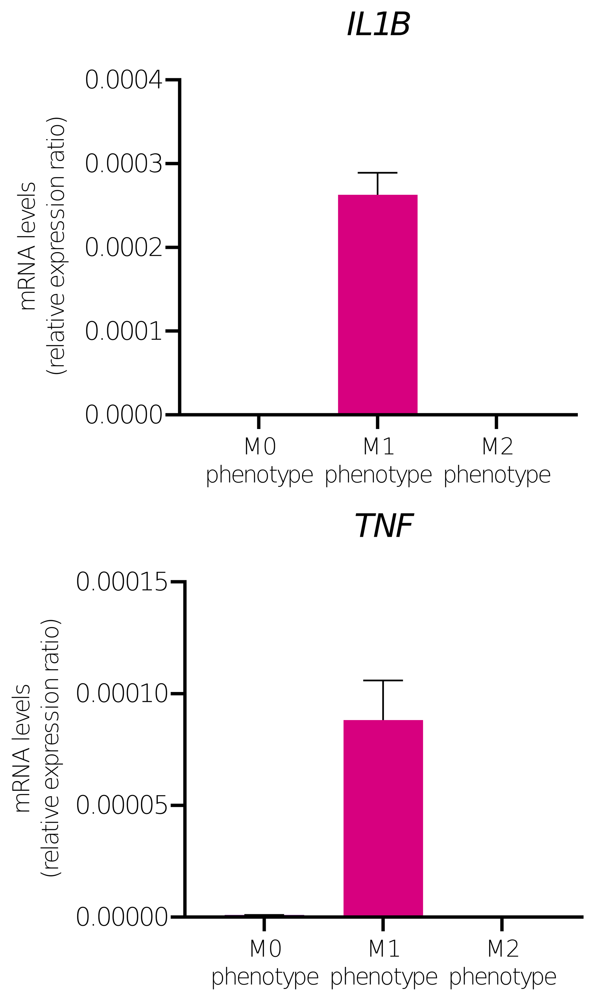
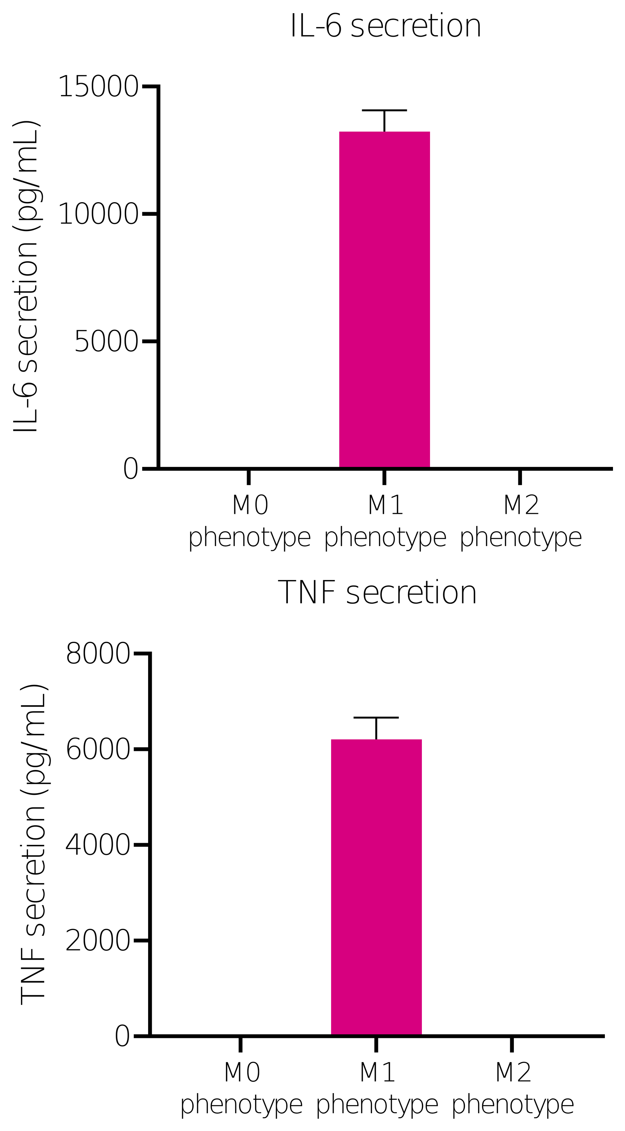
M1 (pro-inflammatory) polarisation: IL1B and TNF transcript induction, with IL-6 and TNF secretion into the media.
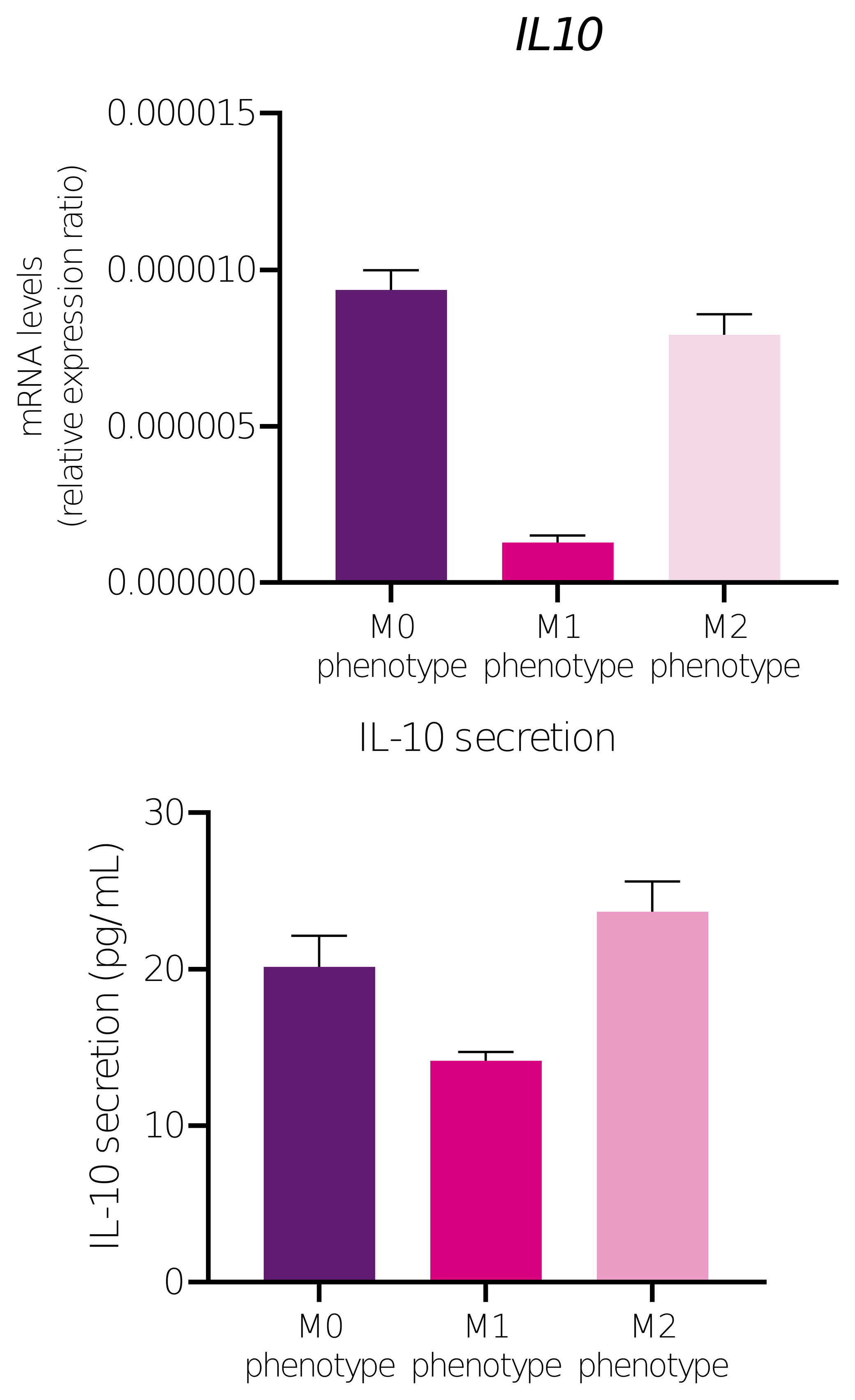
M2 (anti-inflammatory) polarisation: IL10 transcript and secreted protein, restored relative to M1 and comparable to M0.
Direct comparison with THP-1-derived macrophages further highlighted differences in phenotypic responsiveness. Following M2-polarising stimulation, pixMac showed strong induction of MRC1 (CD206), a well-established marker associated with alternatively activated macrophage phenotypes, whereas THP-1 cells showed little or no comparable increase. Baseline lineage-marker expression also differed between the two systems: pixMac expressed CD14, CD68 and MRC1 at readily detectable levels, consistent with a differentiated monocyte/macrophage phenotype, while expression of these markers was minimal or close to background in THP-1 cells.
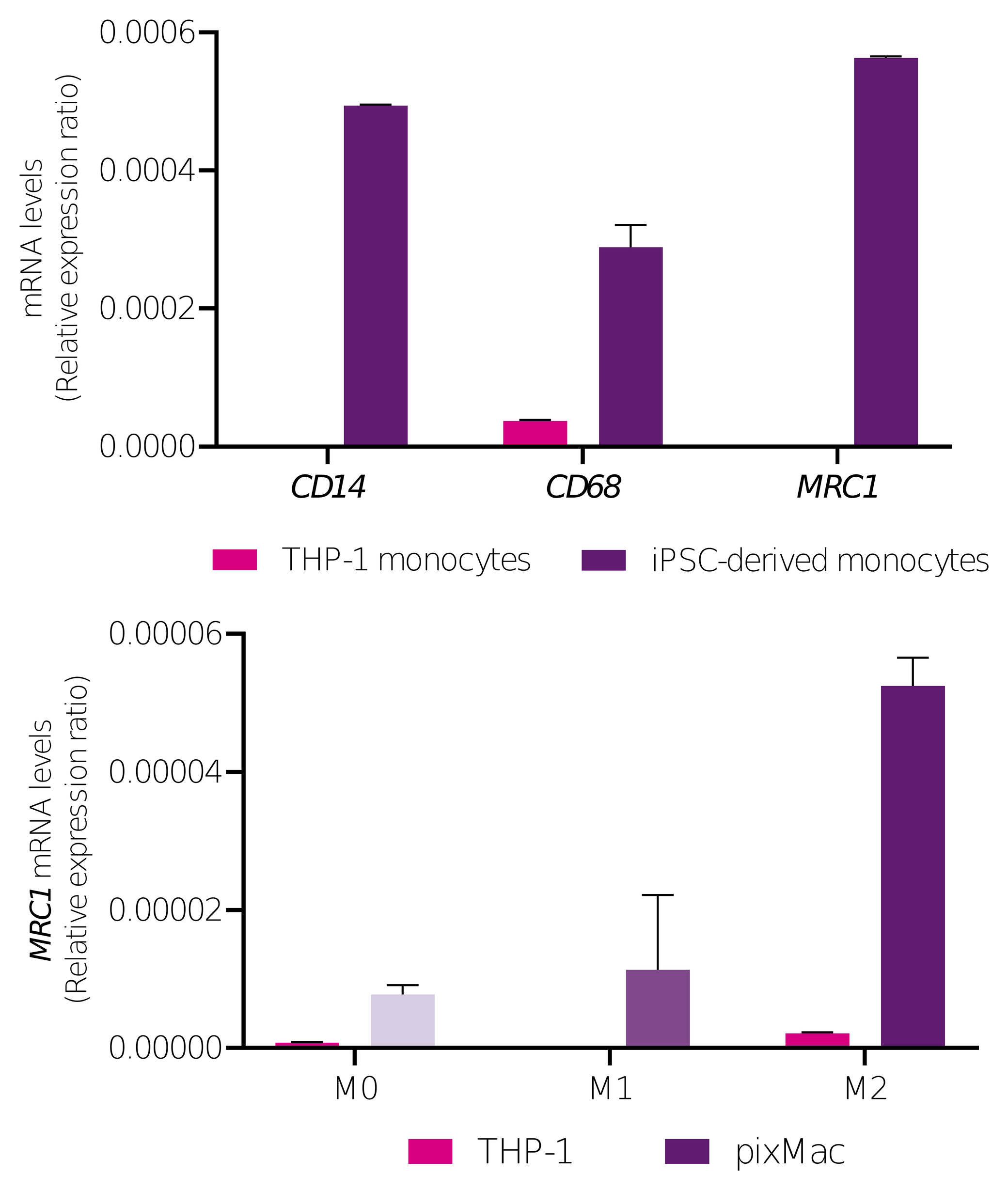
pixMac versus THP-1: authentic monocyte lineage marker expression, CD14/CD68/MRC1 (left), and genuine MRC1 (CD206) induction on M2 polarisation (right). THP-1 is near-negative for lineage markers and fails to mount a comparable M2 response.
Why Does Donor Matching Matter for an Immune Cell Specifically?
Genetic background is particularly important when incorporating immune cells into multicellular in vitro models. Introducing macrophages from one donor background into hepatocyte, stellate cell or organoid systems derived from another can introduce allogeneic immune interactions, including responses driven by differences in HLA genotype. These responses can alter cytokine signalling, immune activation and downstream cellular phenotypes, creating a confounding variable that is unrelated to the compound, disease state or experimental condition under investigation.
pixMac is available on the same donor backgrounds as pixlbio’s pixHep and pixStellate products, enabling the construction of HLA-matched co-culture and tri-culture systems. Using genetically matched cell populations reduces the risk of allogeneic immune activation and allows immune and inflammatory readouts to be interpreted with greater confidence as responses to the experimental perturbation rather than to donor mismatch within the model.
Where Can pixMac Go Beyond the Liver?
pixMac is not restricted to liver-specific applications. As a human macrophage model, it can be incorporated into a range of multicellular systems in which an innate immune component is required. The same donor-matching principle that supports its use with pixHep and pixStellate can also be applied to other tissue models, including intestinal organoids and additional iPSC-derived cell types, enabling immune responses to be studied within a genetically matched cellular context. pixMac can also be used in monoculture to investigate macrophage-specific responses to compounds, including inflammatory activation, immunomodulation and cytokine secretion, independently of other cell types.
pixMac can additionally be generated from customer-provided iPSC lines, enabling the development of HLA-matched macrophages on a defined genetic background. This is particularly relevant for programmes focused on disease-associated variants, donor-specific biology, or population-specific genetic backgrounds, where the availability of a genetically matched immune component can improve the physiological relevance and interpretability of complex in vitro models.
Why Isn't a Hepatocyte Monoculture Enough?
Hepatocyte monocultures capture many important aspects of liver function, but they cannot reproduce the multicellular interactions that drive inflammation and fibrogenesis during chronic liver injury. This is particularly relevant in metabolic dysfunction-associated steatotic liver disease (MASLD), which affects a substantial proportion of the global adult population and can progress from steatosis to steatohepatitis, fibrosis and cirrhosis. Importantly, fibrosis stage is one of the strongest predictors of long-term clinical outcome, with mortality increasing markedly as disease progresses toward cirrhosis.
A similar limitation applies to drug-induced liver injury, where preclinical models do not consistently predict human hepatotoxicity. Several compounds, including tolcapone, ximelagatran and lumiracoxib, progressed through preclinical development before clinically significant liver toxicity became apparent. These limitations reflect, in part, the fact that key pathological responses are not hepatocyte-autonomous. Hepatic stellate cells are central to extracellular matrix deposition and fibrosis, while macrophages regulate inflammatory signalling and can influence both hepatocyte injury and stellate-cell activation. Incorporating these additional cell types therefore enables inflammatory and fibrogenic responses to be measured directly, providing biological information that cannot be obtained from hepatocyte monoculture alone.
What Does a Three-Lineage Liver Model Actually Add?
Each lineage within the tri-culture contributes a distinct component of liver physiology and disease biology. pixHep provides the hepatocellular compartment, supporting metabolic functions including lipid handling, glucose metabolism and fatty-acid oxidation, while also representing a major site of xenobiotic metabolism and drug bioactivation. pixStellate represents the hepatic stellate-cell compartment, which in its quiescent state is associated with vitamin A storage but, following injury-associated activation, can acquire a myofibroblast-like phenotype characterised by increased extracellular matrix and collagen production. pixMac provides the innate immune component, contributing inflammatory mediators such as TNF, IL-1β and CCL2, as well as signalling pathways, including TGF-β and PDGF, that are closely associated with stellate-cell activation and fibrogenesis.
The value of the tri-culture lies in the interaction between these compartments. Macrophage-derived inflammatory and profibrogenic signals can promote stellate-cell activation, while activated stellate cells alter the extracellular matrix and secrete mediators that can, in turn, influence macrophage behaviour. These reciprocal interactions are important features of progressive liver injury and cannot be reproduced in hepatocyte monoculture alone.
There is also increasing evidence that macrophage phenotype is strongly influenced by the local tissue microenvironment. Published studies have shown that direct co-culture of iPSC-derived macrophages with iPSC-derived hepatocytes can promote acquisition of Kupffer-cell-like characteristics while simultaneously supporting hepatocyte maturation, including reduced expression of fetal markers and increased expression of cytochrome P450 genes. Importantly, some of these effects depend on direct cellular interactions and are not fully reproduced by hepatocyte-conditioned medium alone.
For this reason, co-seeding the three cell populations provides a model in which hepatocytes, stellate cells and macrophages can establish reciprocal signalling from the outset, rather than introducing the immune compartment only after the other populations have established independently. This approach is intended to capture contact-dependent and paracrine interactions that may contribute to inflammatory, fibrogenic and immune-mediated drug responses and that are absent from simpler hepatocyte-only or sequentially assembled systems.
Is the Tri-Culture Actually a Tri-Culture?
Molecular and immunocytochemical analyses confirm both the maintenance of the three cellular compartments and the emergence of inflammatory and fibrogenic phenotypes within the tri-culture. Relative to pixHep monoculture, the tri-culture shows increased expression of stellate-associated fibrogenic markers, including ACTA2 and COL1A1, together with macrophage-associated inflammatory markers such as CD68 and TNF, indicating activation of biological pathways that cannot be captured in a hepatocyte-only system. In parallel, immunocytochemistry confirms retention of the individual cell identities within the multicellular model, with albumin identifying the hepatocyte compartment, GFAP the stellate-cell compartment and IBA1 the macrophage compartment, demonstrating that the three lineages remain phenotypically distinguishable while co-existing within the same culture system.
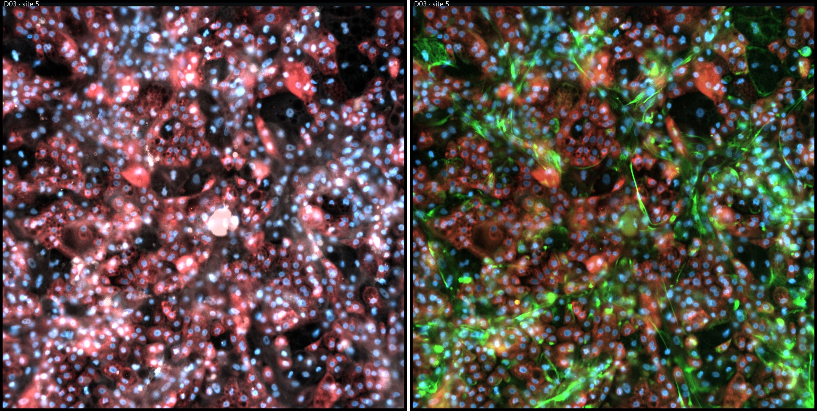
Immunocytochemistry of pixHep/pixMac (left) and pixHep/pixStellate (right) pairings within the tri-culture. Albumin (hepatocyte), GFAP (stellate), IBA1 (mature macrophage).
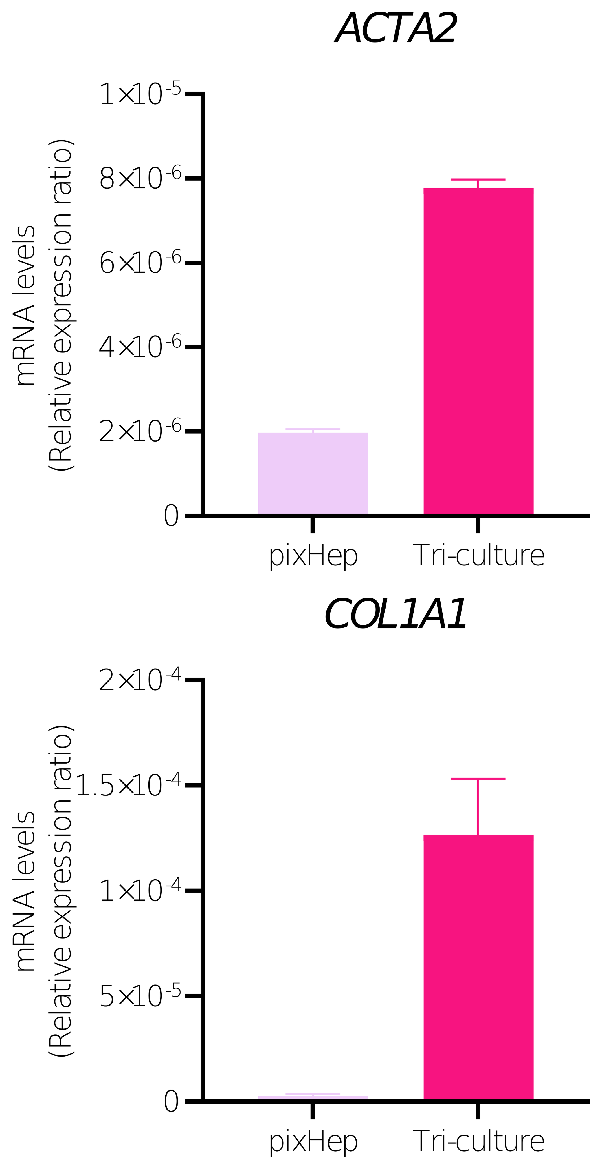
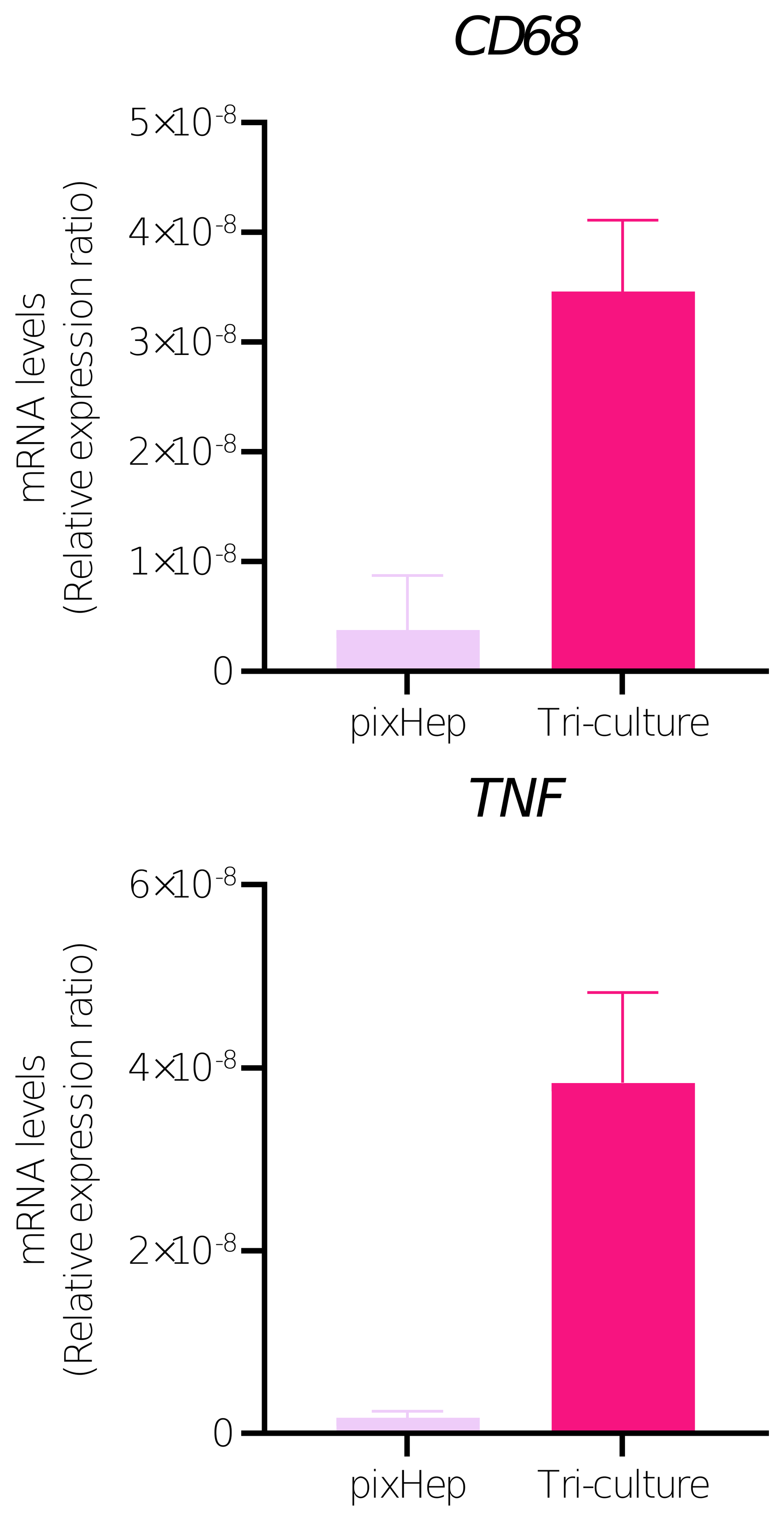
Stellate markers (ACTA2, COL1A1) and macrophage markers (CD68, TNF) in tri-culture versus pixHep monoculture.
Can the Tri-Culture Separate a Toxic Drug from a Safe One?
A key test of the model is its ability to distinguish compounds with similar pharmacological activity but different hepatotoxicity profiles. Tolcapone and entacapone are both catechol-O-methyltransferase inhibitors used in the treatment of Parkinson’s disease, but differ substantially in their clinical liver-safety profiles, with tolcapone associated with clinically significant hepatotoxicity whereas entacapone has a considerably lower hepatic liability.
Within the tri-culture, CYP3A4 expression was increased relative to pixHep monoculture, consistent with enhanced hepatocyte maturation in the multicellular environment. Tolcapone reduced this increase in CYP3A4 expression, whereas entacapone did not produce a comparable effect. The two compounds were also differentiated at the immune level: tolcapone increased TNF expression and was associated with increased secretion of TNF protein, while entacapone induced little to no detectable TNF secretion. This inflammatory response is inherently inaccessible in hepatocyte monoculture because the relevant immune compartment is absent. Pro-collagen I alpha 1 secretion provided an additional stellate-cell-derived readout that was measurable only in the multicellular system and was differentially modulated following compound exposure. Together, these responses demonstrate that the tri-culture can resolve hepatocellular, inflammatory and fibrogenic effects within the same experimental system.
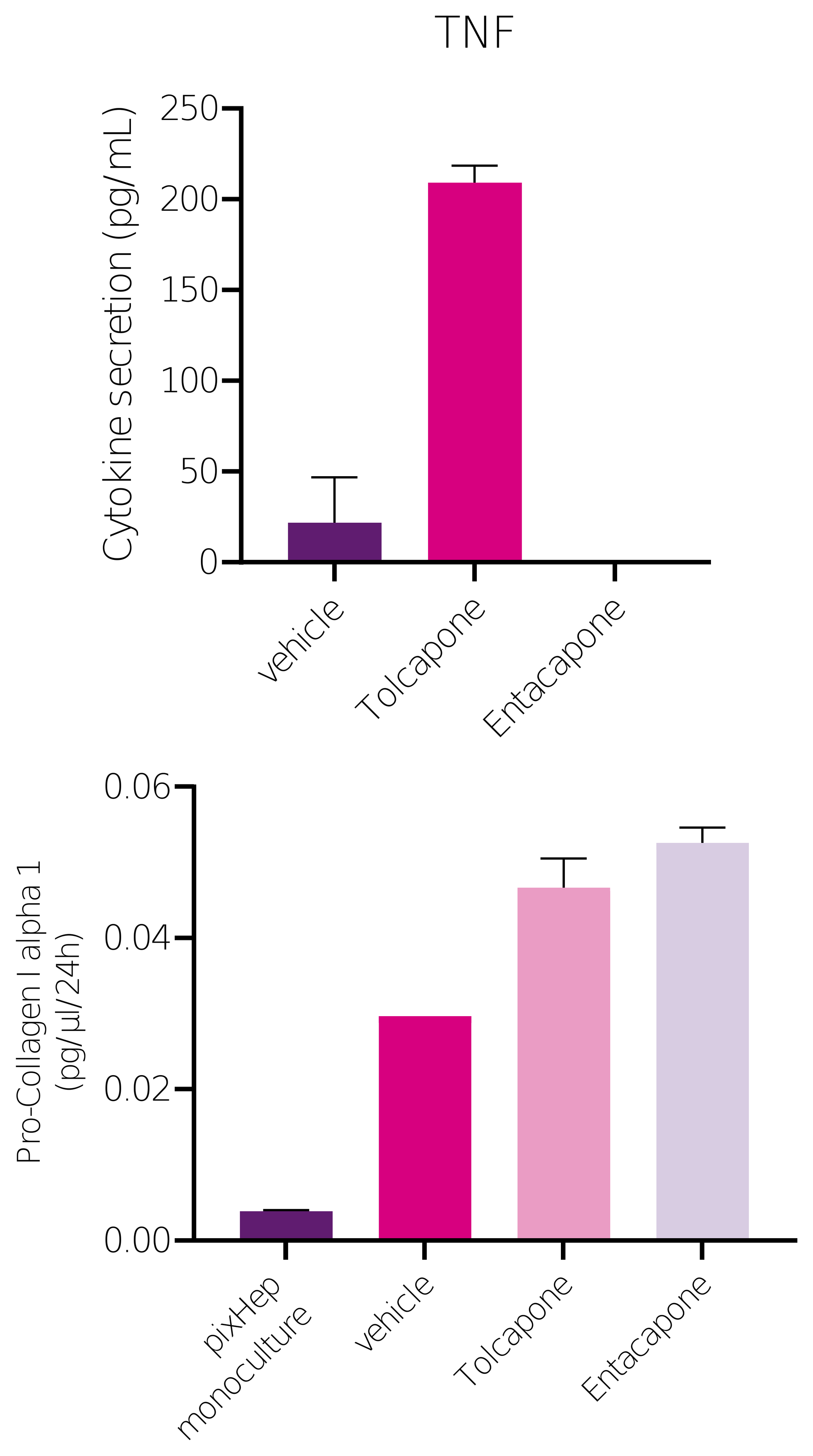
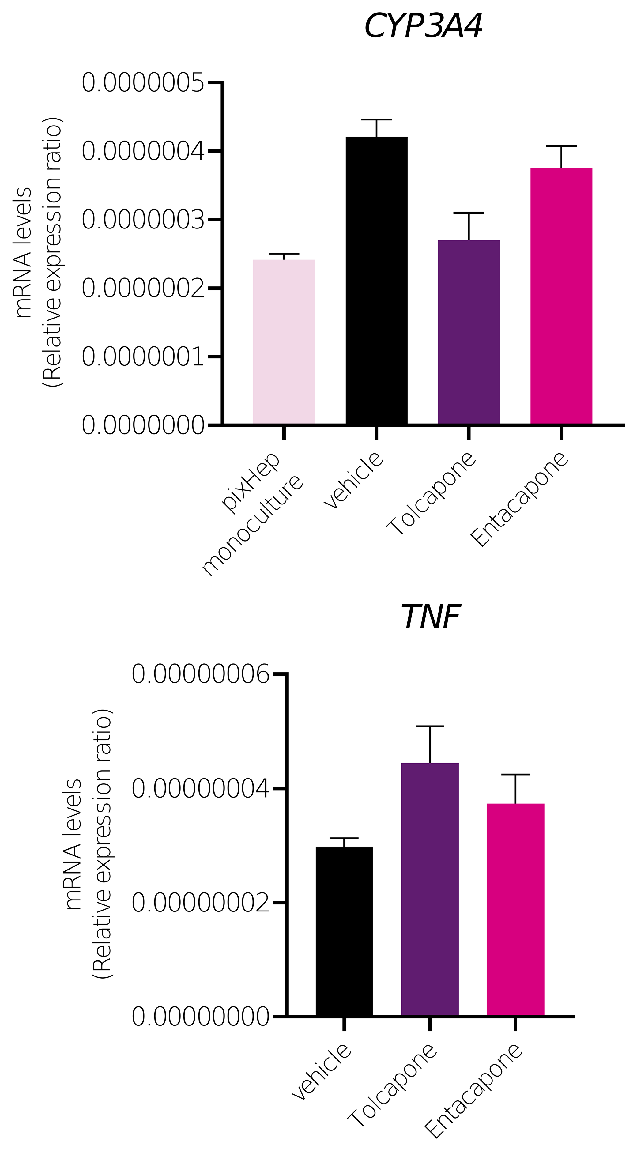
Tri-culture separates hepatotoxic tolcapone from non-hepatotoxic entacapone on CYP3A4 maturation, TNF transcript and secretion, and pro-collagen I alpha 1, vehicle vs. tolcapone vs. entacapone.
Does the Tri-Culture Respond to Metabolic Disease Conditions Too?
The tri-culture also responds to a MASH-relevant metabolic challenge. Following five days of exposure to free fatty acids, expression of CPT1A and G6PC increased by approximately two-fold, indicating substantial adaptation of pathways involved in fatty-acid oxidation and hepatic glucose metabolism. These metabolic changes were accompanied by increased expression of CD68 and TNF, together with an approximately 20% increase in pro-collagen I alpha 1 secretion relative to the control-diet condition.
Importantly, these inflammatory and fibrogenic responses developed in the absence of exogenous inflammatory cytokines or direct profibrogenic stimulation. The data therefore indicate that metabolic stress alone is sufficient to initiate coordinated metabolic, inflammatory and fibrogenic responses within the tri-culture, recapitulating key features associated with the progression of MASH.
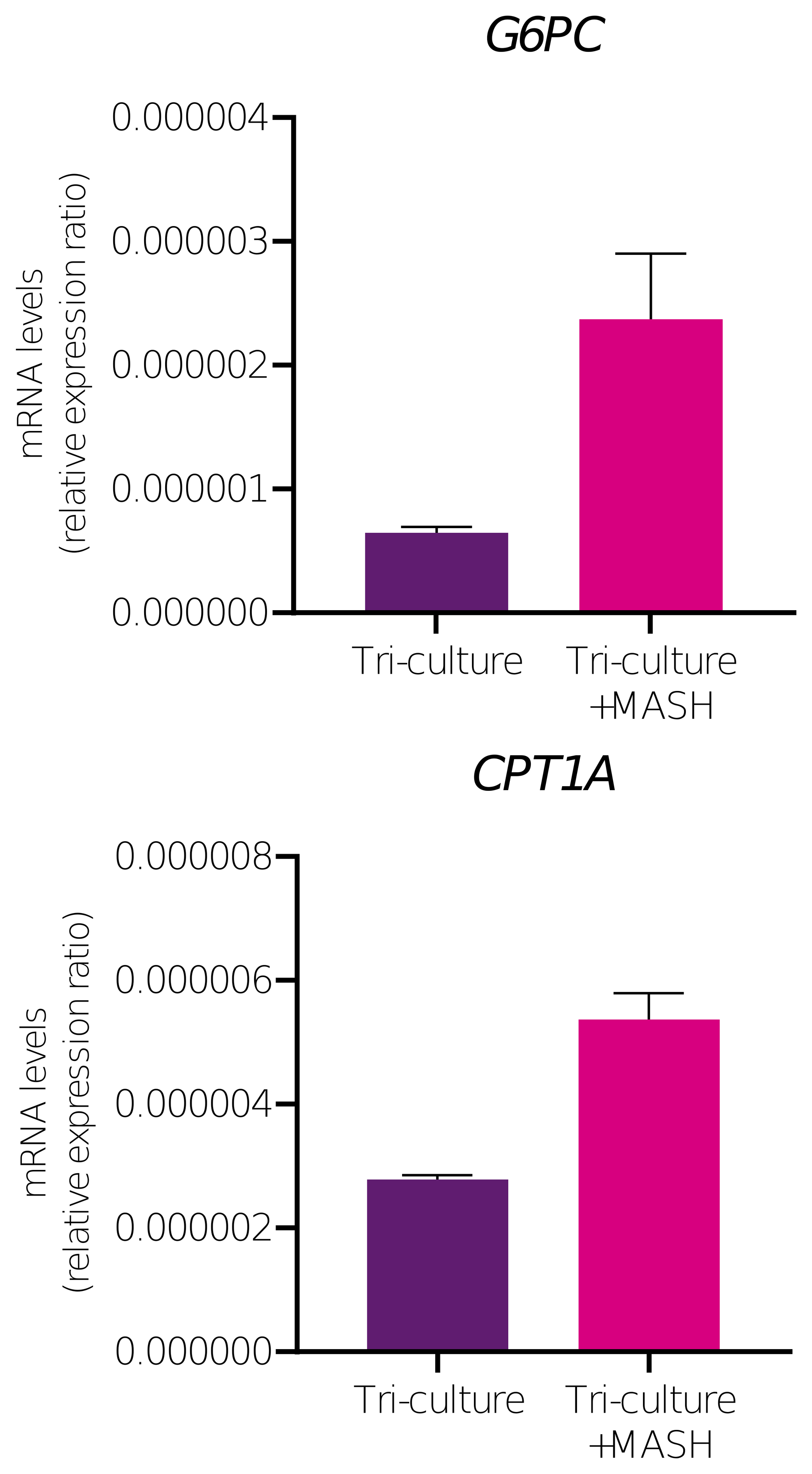
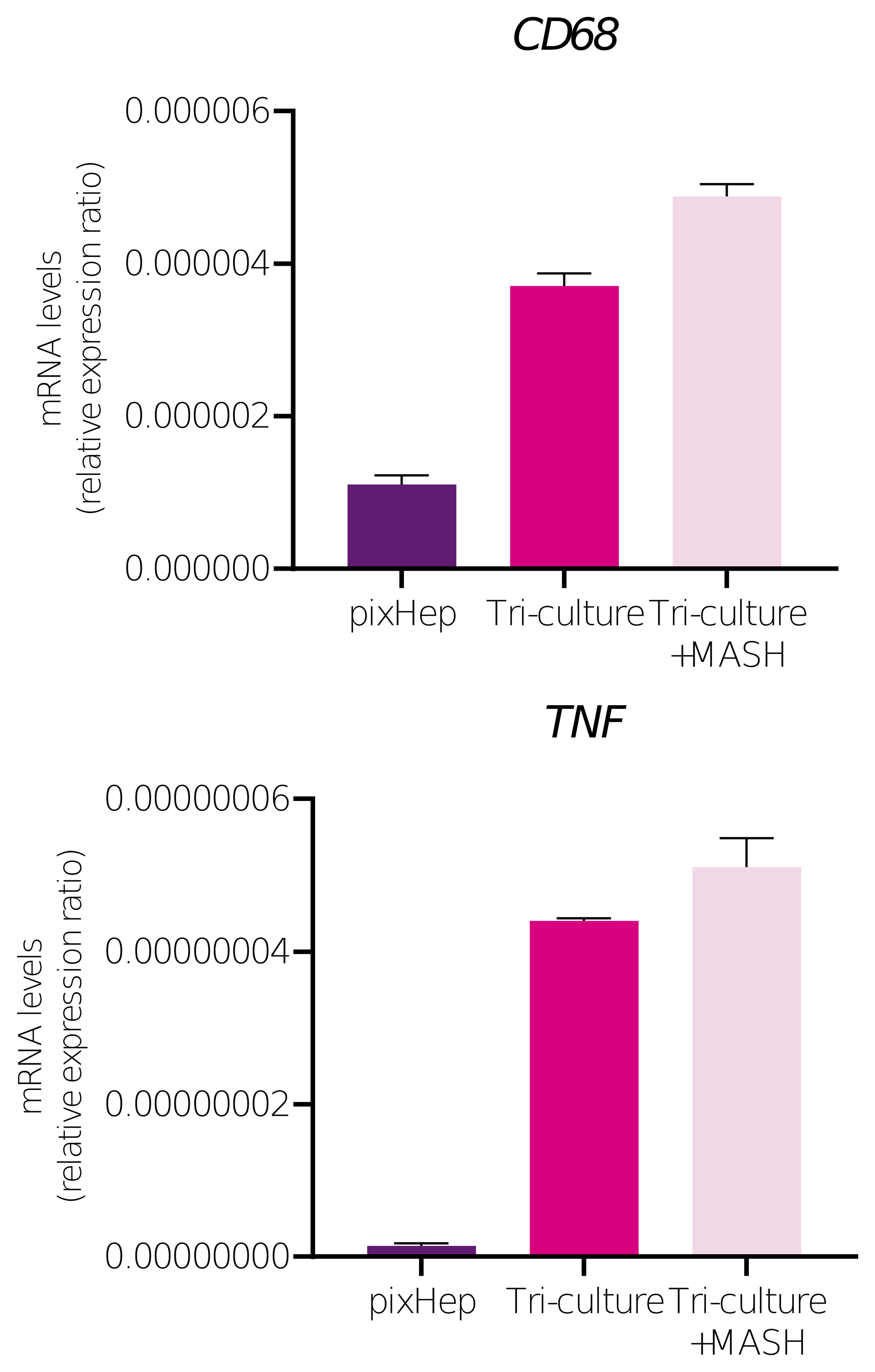
Compensatory lipid-handling genes (G6PC, CPT1A) and inflammatory markers (CD68, TNF) in tri-culture following 5-day MASH-diet challenge.
What's Next: pixCellPaint on the Tri-Culture
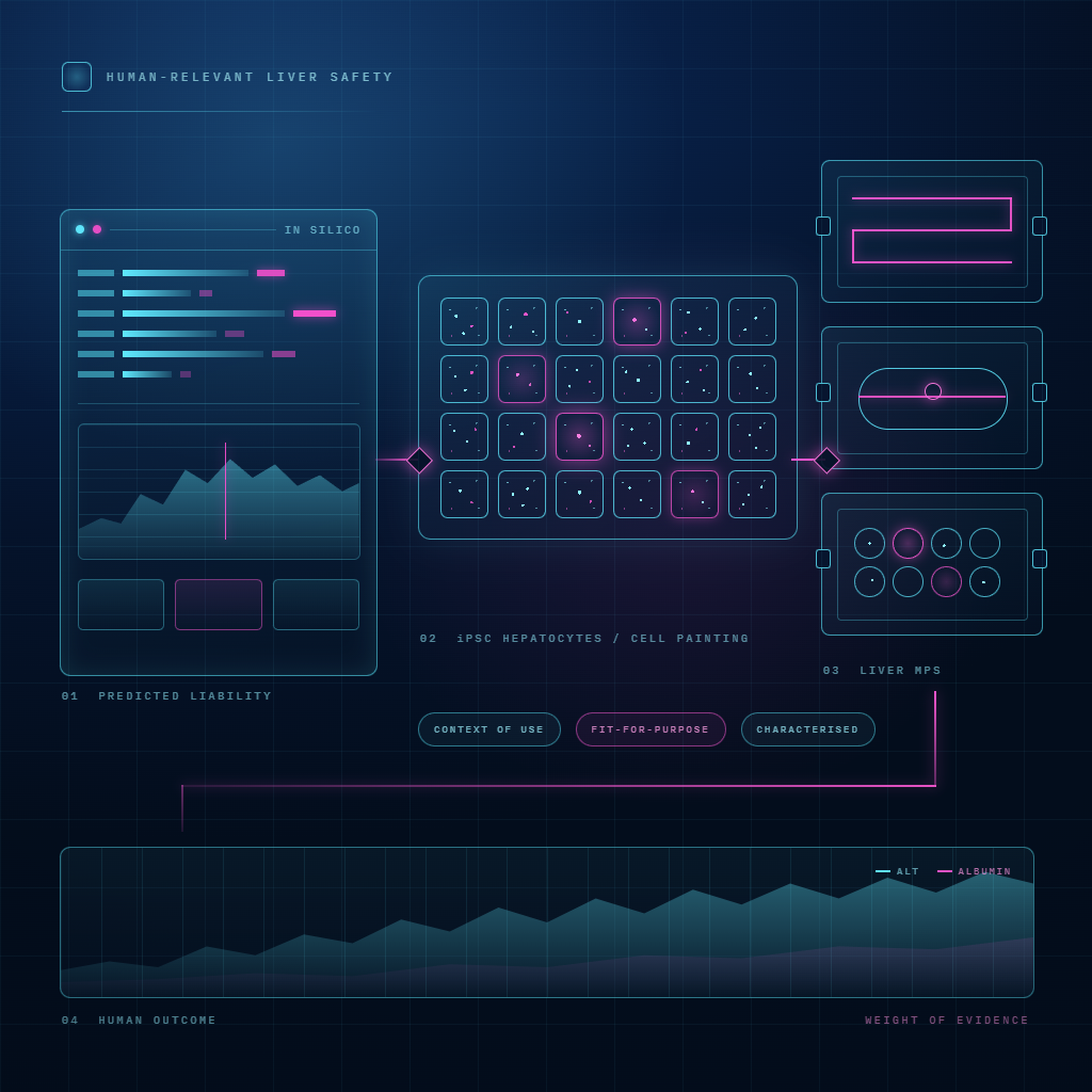
The functional endpoints above already show the tri-culture can separate a toxic drug from a safe one and respond to a metabolic disease challenge. The natural next step is phenotypic profiling on top of that functional foundation rather than in place of it. Three interacting cell populations are exactly where an unbiased, high-content readout earns its place: a handful of pre-selected markers can only capture a fraction of what is actually changing across three cell types at once. pixlbio's own view, based on internal experience with Cell Painting performance across dimensionalities, is that this tri-culture is better suited to 3D profiling than the standard 2D format, and that work is what comes next for this model.
Conclusion
Human iPSC-derived macrophages for unraveling human macrophage biology. PMC
Induced pluripotent stem cell-derived macrophages enable broad modeling of human inflammasome signaling. PMC
Nascimento CR, Fernandes NAR, Gonzalez Maldonado LA, Rossa Junior C (2022). Comparison of monocytic cell lines U937 and THP-1 as macrophage models for in vitro studies. Biochemistry and Biophysics Reports
The Role of Kupffer Cells and Liver Macrophages in the Pathogenesis of Metabolic Dysfunction-Associated Steatotic Liver Disease (2026). Biomolecules, MDPI
Macrophages and Kupffer Cells in Drug-Induced Liver Injury. ScienceDirect
Krenkel O, Tacke F (2017). Liver macrophages in tissue homeostasis and disease. Nature Reviews Immunology
Lee CZW, Tasnim F, Huang X, et al. (2026). Direct contact between iPSC-derived macrophages and hepatocytes drives reciprocal acquisition of Kupffer cell identity and hepatocyte maturation. eLife
Tolcapone. LiverTox: Clinical and Research Information on Drug-Induced Liver Injury, NIDDK
Miao L, Targher G, Byrne CD, et al. (2024). Current status and future trends of the global burden of MASLD. Trends in Endocrinology and Metabolism
Ekstedt M, Hagström H, Nasr P, et al. (2015). Fibrosis stage is the strongest predictor for disease-specific mortality in NAFLD after up to 33 years of follow-up. Hepatology
pixlbio. Human iPSC Disease Models for Drug Discovery. pixlbio Disease Models
pixMac is available individually or donor-matched with pixHep and pixStellate for co-culture and tri-culture work, and as a custom HLA-matched service against a customer's own iPSC line.
To discuss pixMac for your programme, book a call with our team.
Tags: pixMac · iPSC-Derived Macrophages · Kupffer Cells · Tri-Culture · Immunology · Liver Disease · DILI · MASLD · Cell Painting · Drug Discovery