No items found.
No items found.

Catalog Numbers: HEP-003, HEP-059, HEP-004, HEP-003-TM6SF2-a, HEP-003-PNPLA3-h

MASLD (Metabolic Dysfunction-Associated Steatotic Liver Disease) Models

iPSC-derived hepatocyte models replicating steatosis, insulin resistance, and inflammation.

  • Cryopreserved vials (1 × 10⁶ viable cells per vial) from defined donor backgrounds, PNPLA3, TM6SF2, and multiple wild-type backgrounds available
  • Inducible steatosis and lipid accumulation validated by BODIPY and cytokine readouts
  • Suitable for mechanistic and compound screening in metabolic disease pathways
Place your Order
Item
pixHep-003 iPSC-derived Hepatocyte Donor 1, Male (Wild Type)
Price
£ 1,700 
0

Our MASLD pixHep™ model replicates the lipid accumulation, metabolic dysfunction, and inflammatory signalling of clinical steatosis.

Available in wild-type or genetic risk backgrounds, it provides a robust platform for screening metabolic modulators, anti-inflammatories, and disease progression blockers.

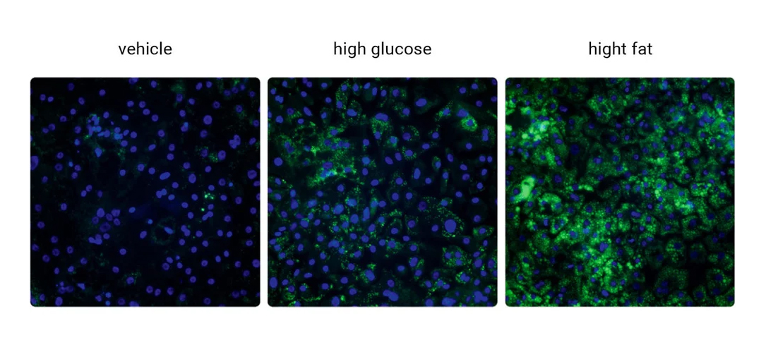
Lipid droplet accumulation confirmed by imaging.

Altered lipid metabolism and oxidative stress markers.

Compatible with CRISPR-driven genetic risk variants.

Co-culture ready for MASH progression modelling.

Technical Data & Functional Validation

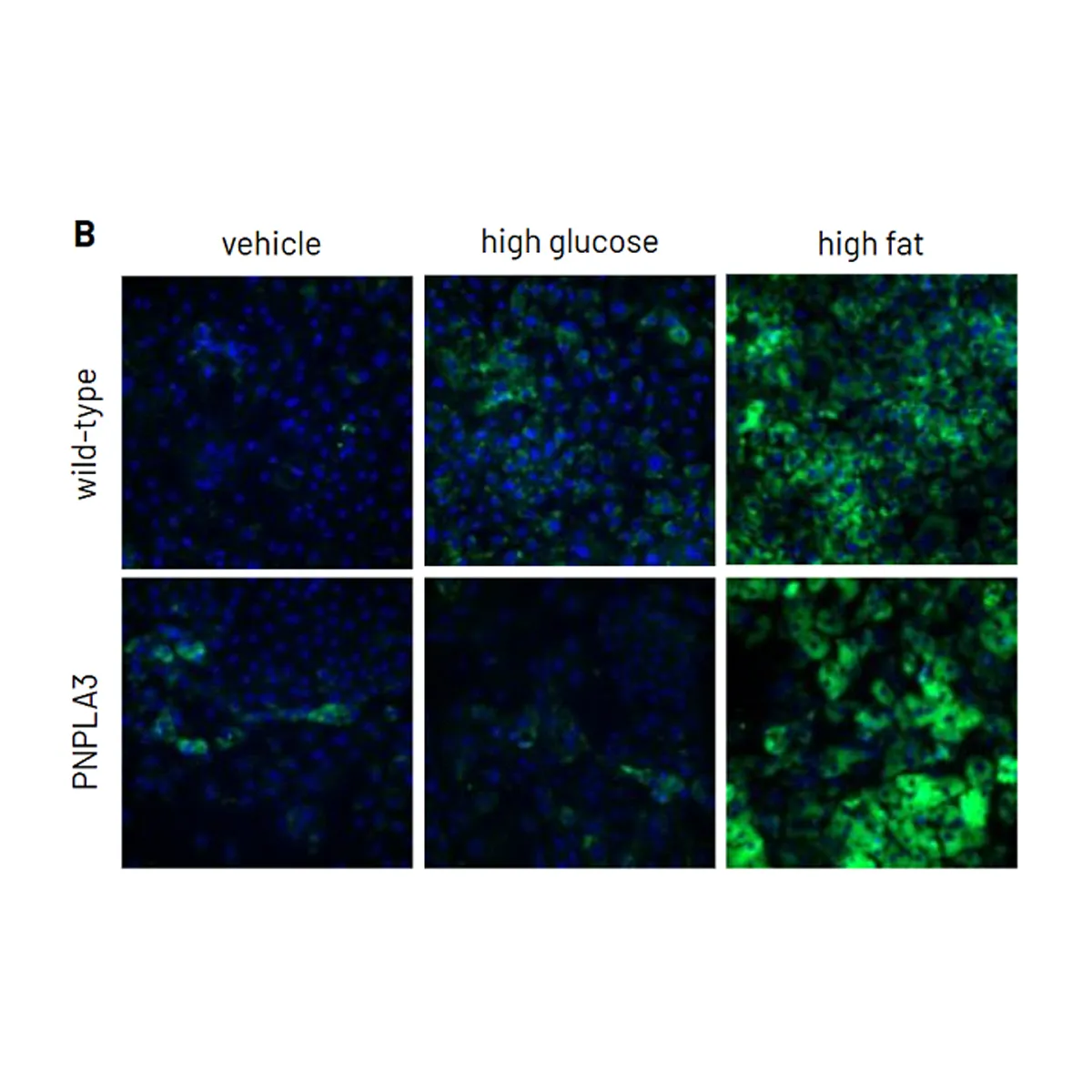
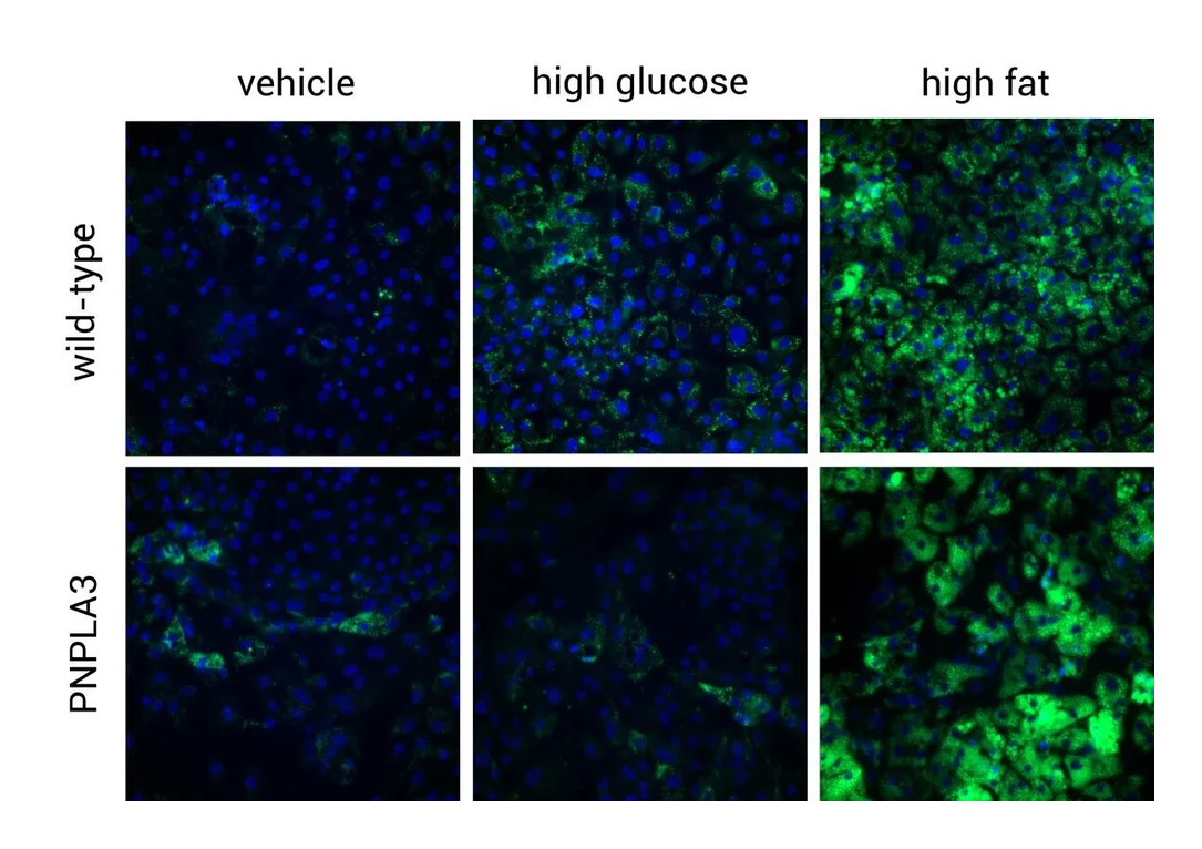
Increased Lipid Accumulation
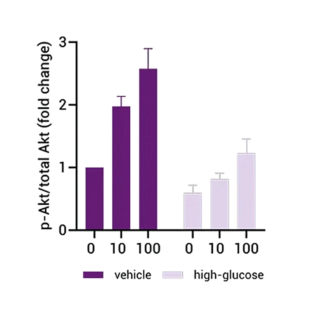
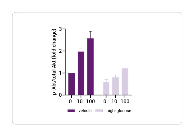
MASLD iPSC hepatocyte model showing lipid droplet accumulation
pixHep MASLD hepatocyte increased lipid accumulation BODIPY staining fluorescence microscopy after 5 days glucose and free fatty acid treatment
pixlbio pixHep demonstrate increased lipid accumulation following 5 days of treatment with glucose and free fatty acids as measured by BODIPY staining. mRNA data were normalized to housekeeping gene PPIA and fluorescence intensity to total nuclei number. All data are presented as mean±SEM of n=3 independent experiments.

Insulin Resistance

pixHep MASLD hepatocytes showing reduced AKT phosphorylation following insulin stimulation 10-100nM indicating hepatic insulin resistance, normalized to total AKT and beta-actin
High-glucose-treated pixlbio pixHeps demonstrate reduced phosphorylation of AKT following stimulation with increasing concentrations of insulin (10-100nM) compared to vehicle-treated cells, indicative of hepatic insulin resistance. Protein data were normalized to total AKT and beta-actin and are presented as mean±SEM of n=3 independent experiments.

Lipotoxicity and Pro-Inflammation

pixHep MASLD hepatocytes showing reduced cell viability and increased pro-inflammatory cytokines IL-1β IL-6 IL-8 TNFα expression following high-fat treatment, mRNA normalized to 18S rRNA
Left: High-fat-treated pixlbio pixHeps demonstrate reduced cell viability compared to vehicle-treated cells, indicative of enhanced lipotoxicity. Right: High-fat-treated pixlbio pixHeps demonstrate increased expression of pro-inflammatory cytokine markers (IL-1β, IL-6, IL-8, TNFα) compared to vehicle-treated cells. mRNA data were normalized to 18S rRNA. All data are presented as mean±SEM of n=2-3 independent experiments.

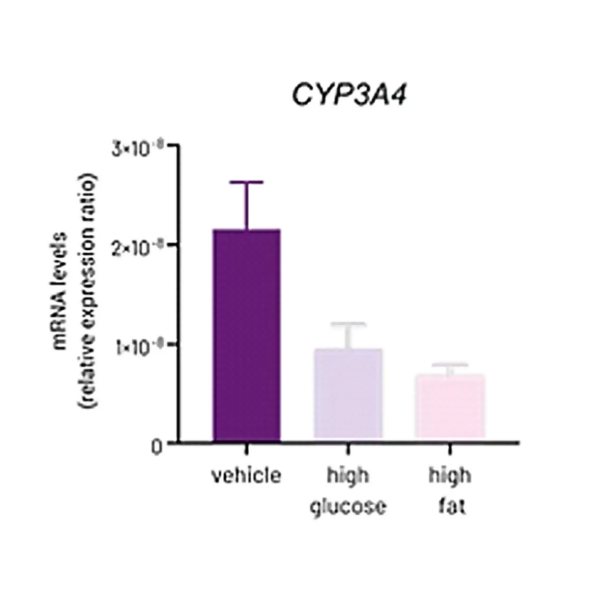
Reduced Phase 1 Activity

pixHep CYP450 gene expression CYP3A4 CYP2B6 CYP2C9 CYP2A6 CYP2C19 CYP2J2 in vehicle high-glucose and high-fat treated hepatocytes, mRNA normalized to 18S rRNA
mRNA expression levels of the Phase 1 CYP450 genes CYP3A4, CYP2B6, CYP2C9, CYP2A6, CYP2C19, and CYP2J2 in vehicle-, high-glucose-, and high-fat treated pixHeps mRNA data were normalized to 18S rRNA and are presented as mean±SEM of n=2-3 independent experiments.

PNPLA3 Variant MASLD Phenotype

PNPLA3-mutant pixHep hepatocytes showing impaired AKT phosphorylation response to insulin 10-100nM compared to wild-type pixHep
AKT phosphorylation in PNPLA3-mutant pixHep following stimulation with increasing concentrations of insulin (10-100nM), revealing impaired response to insulin compared to wild-type pixHep.
Streamline Your Research

Ready to Turn Your Cells Into Data?

Our pixCell portfolio seamlessly integrates into custom data generation projects—from functional assays (pixCellServices) to phenomic analysis (pixCellPaint)—delivering ready-to-use data at your fingertips.