

No items found.
No items found.
Catalog Number: HSC-003
pixStellate iPSC-derived Stellate Cells
Human iPSC-derived stellate cells (1 × 10⁶ viable cells per vial) for modeling fibrosis and hepatic inflammation.
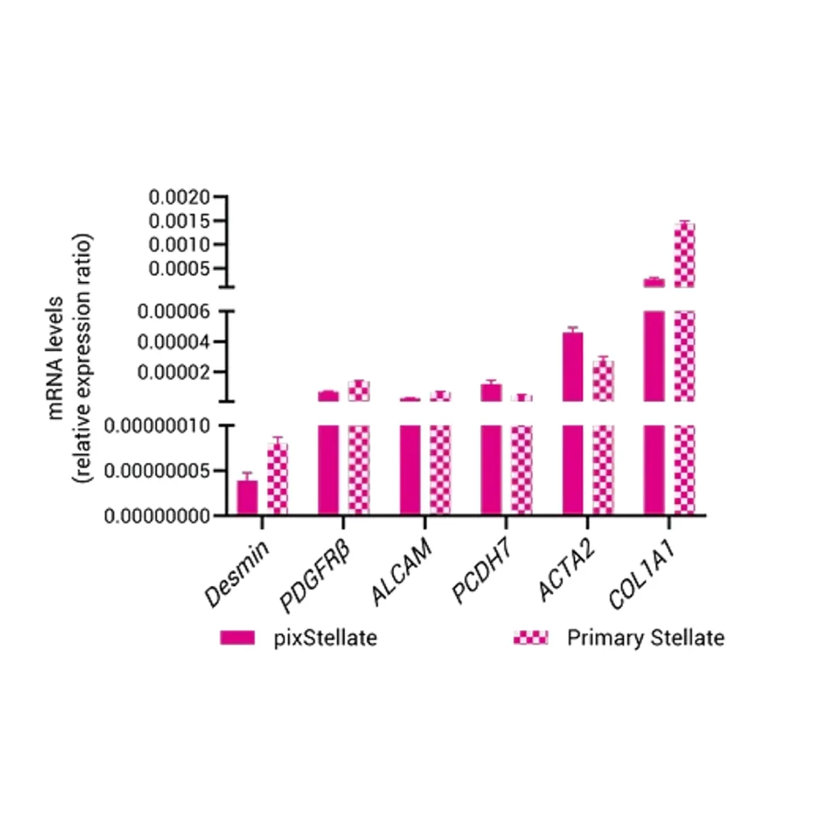
- Express key fibrotic and activation markers (COL1A1, ACTA2, PDGFRβ)
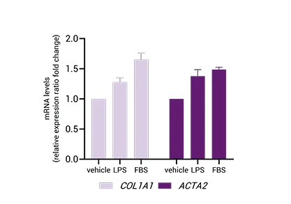
- Respond to TGF-β and LPS stimulation for fibrosis induction studies
- Donor-matched Co-culture compatible with pixHep for MASLD and steatohepatitis modelling
Place your Order
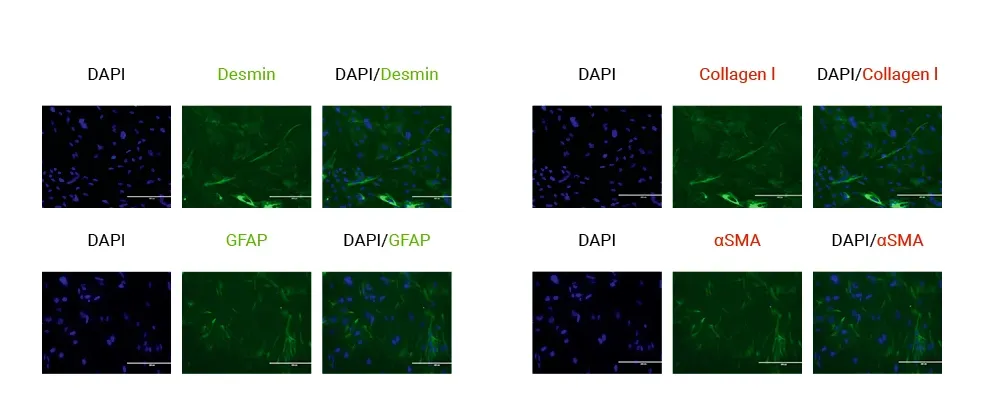
pixStellate™ are iPSC-derived stellate cells that maintain hallmark markers (α-SMA, desmin, GFAP) and activate under fibrogenic stimuli.
They can be used alone or paired with pixHep™ to model fibrosis progression, MASLD-to-NASH transitions, or chronic liver injury.
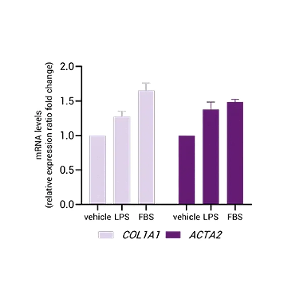
Quiescent in baseline culture; responsive to injury cues.
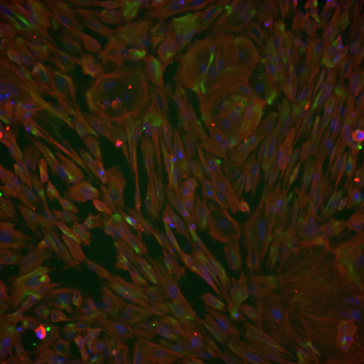
Express hallmark stellate markers confirmed via immunostaining.
Upregulate pro-fibrotic genes upon activation.
Available from healthy or disease donor lines.
Compatible with custom genetic engineering
Technical Data & Functional Validation
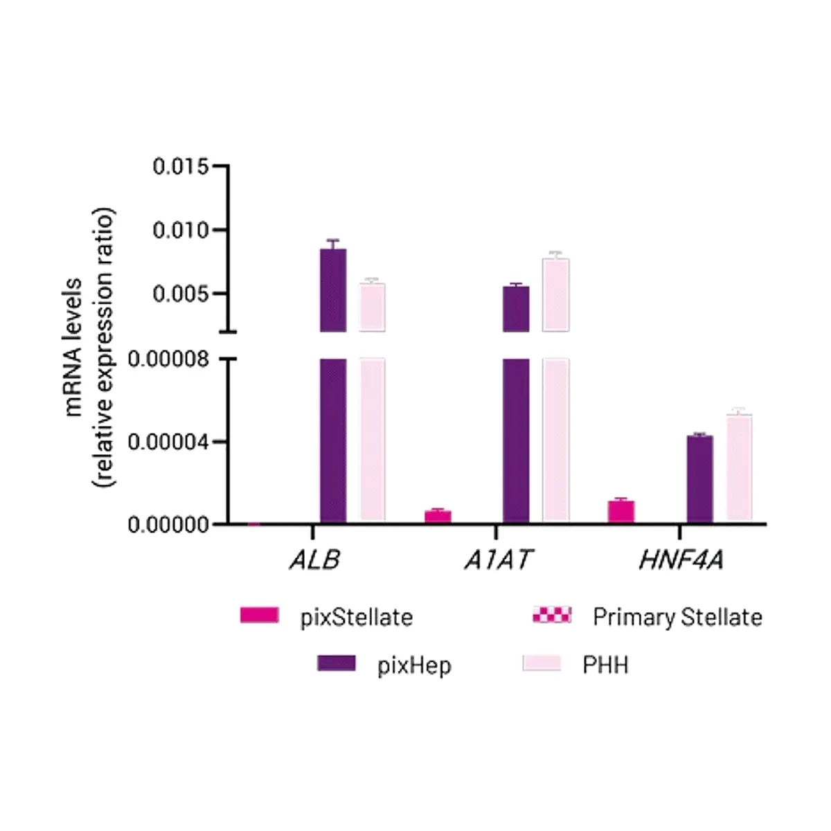
pixlbio iPSC-derived Hepatic Stellate Cells (pixStellate) express key HSC markers at gene and protein level
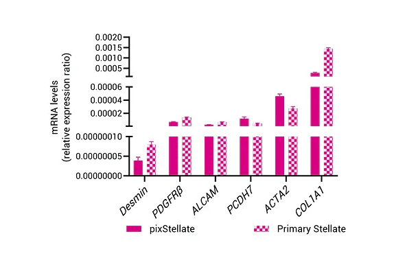
Key HSC Markers (mRNA)

Key HSC Markers (Protein)
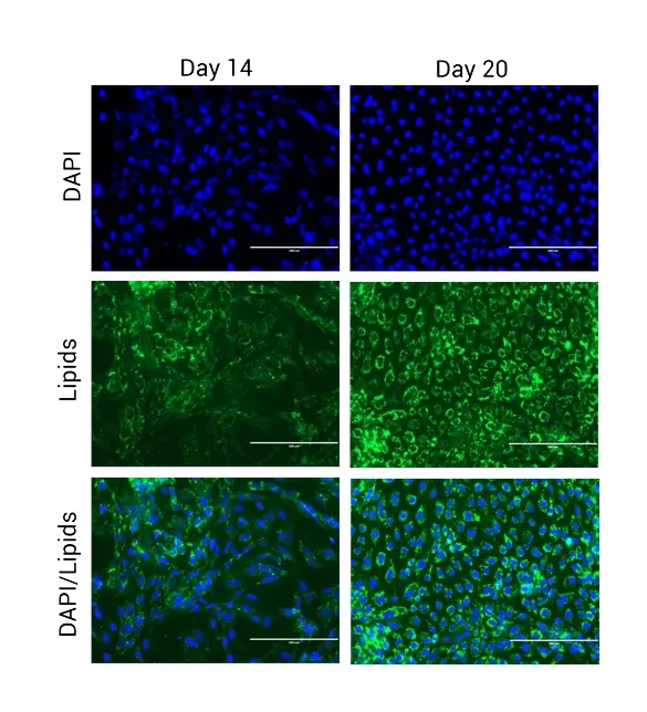
Quiescent Phenotype
pixStellate Activation


Streamline Your Research
Ready to Turn Your Cells Into Data?
Our pixCell portfolio seamlessly integrates into custom data generation projects—from functional assays (pixCellServices) to phenomic analysis (pixCellPaint)—delivering ready-to-use data at your fingertips.

