
No items found.
No items found.
Catalog Numbers: HEP-001, HEP-003-AAT-z, HEP-003-AAT-a
pixHep A1ATD (Alpha-1 Antitrypsin Deficiency) Models
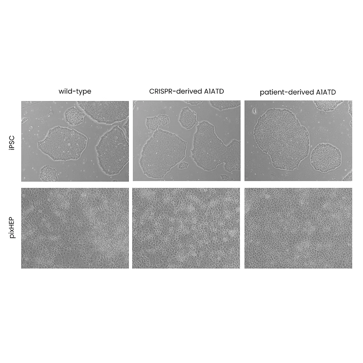
pixHep iPSC-derived hepatocyte models representing patient-derived and CRISPR-engineered genotypes of SERPINA1-E342K (PiZZ and PiMZ).
- Cryopreserved vials (1 × 10⁶ viable cells per vial) available as patient-derived PiZZ, CRISPR PiZZ (homozygous), and CRISPR PiMZ (heterozygous) lines
- Reproduce A1AT misfolding, aggregation, and intracellular inclusion formation
- Suitable for proteostasis and autophagy pathway analysis, therapeutic rescue screening, and comparative mechanism-of-action studies
Place your Order
Our A1ATD pixHep™ model carries the SERPINA1 Z mutation, producing misfolded A1AT protein and accumulation within hepatocytes.
It is ideal for studying ER stress, protein aggregation, and therapeutic rescue strategies.
Validated A1AT misfolding and accumulation phenotype.
Quantifiable ER stress and apoptosis markers.
Compatible with gene therapy and proteostasis-targeting compounds.
Available with isogenic wild-type controls.
Technical Data & Functional Validation
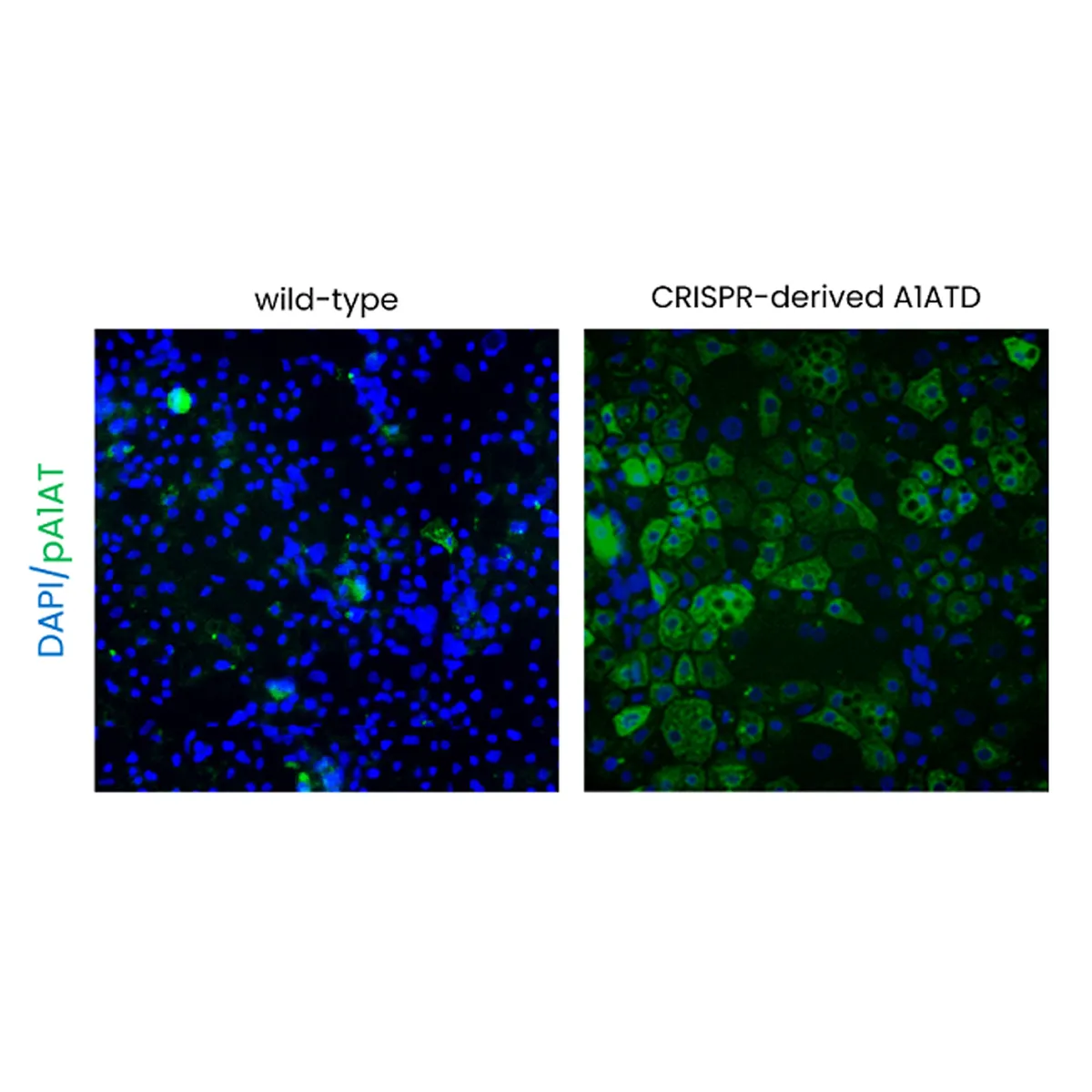
CRISPR Derived Model

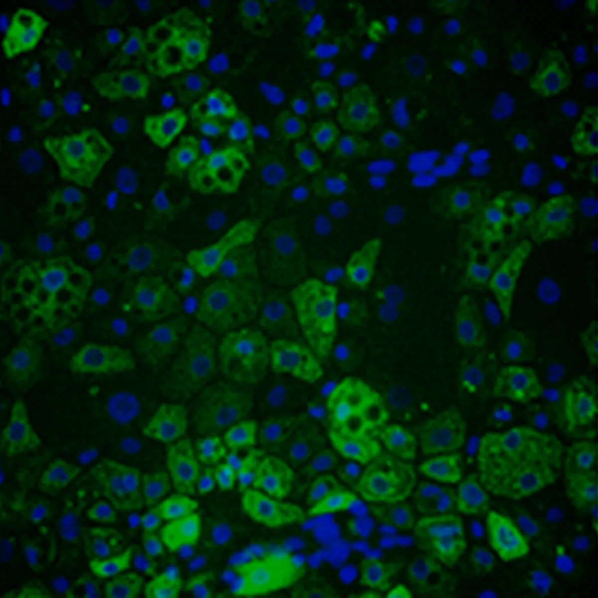
Phenotypic Validation of Polymeric A1AT (ZZ)
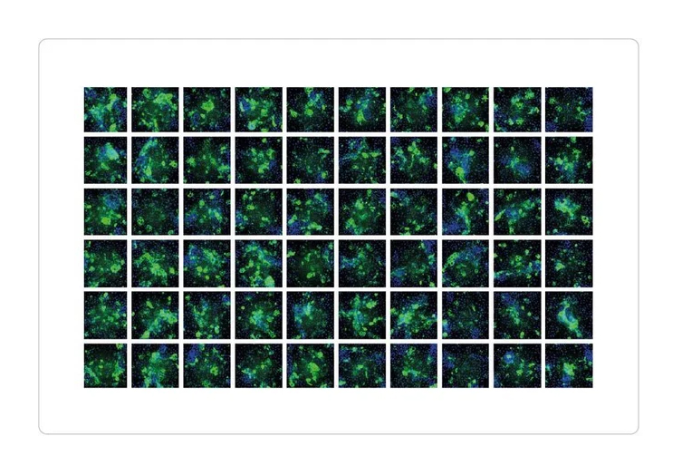
Intracellular of polymeric A1AT in ZZ-hepatocytes

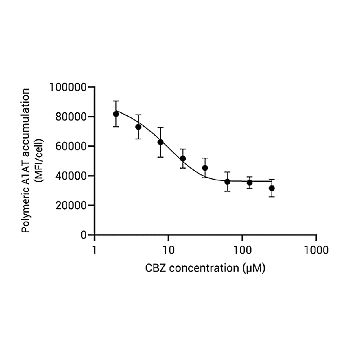
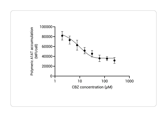
Decrease of intracellular polymeric A1ATD
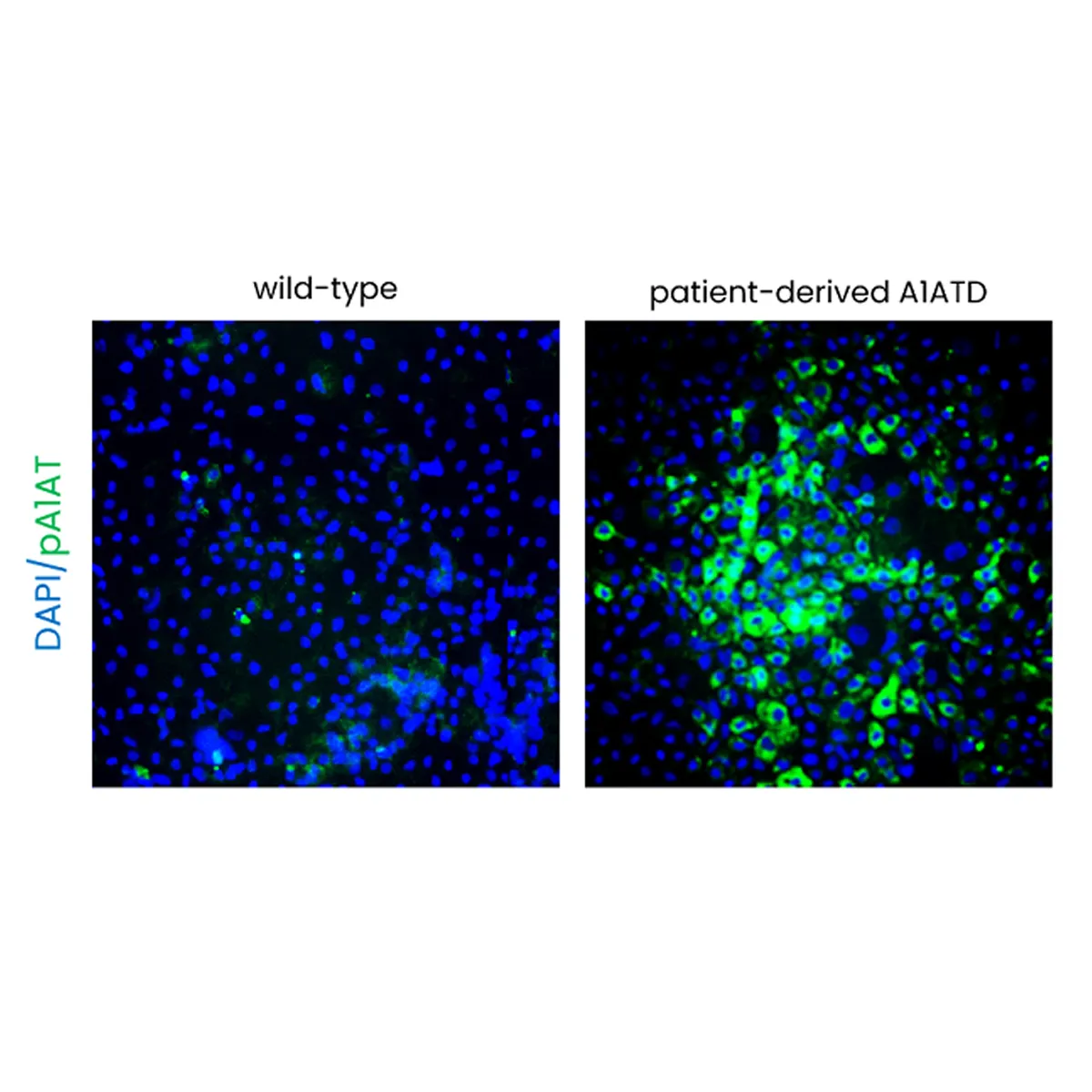
Patient-derived A1AT Accumulation

Donor dependent A1AT Acumulation

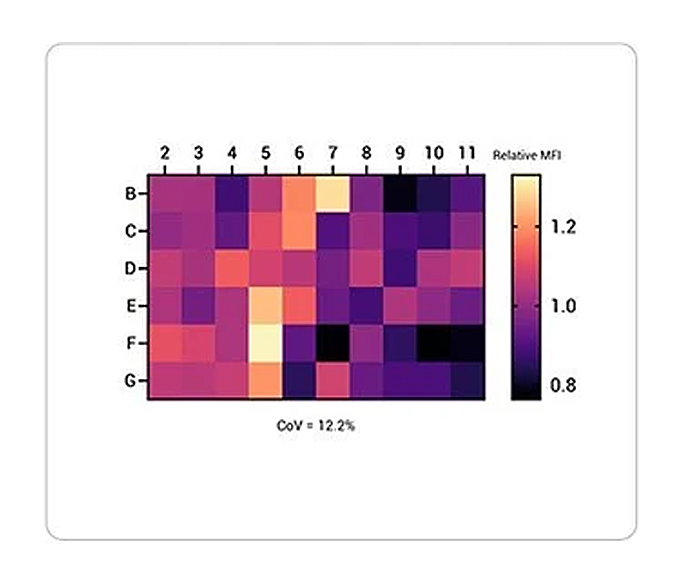
Below: Quantification of the intracellular levels of polymeric A1AT in pixlbio A1ATD hepatocytes across the central wells of a 96 well plate (n=3).
Patient-derived pixHeps respond to Carbamazepine


Streamline Your Research
Ready to Turn Your Cells Into Data?
Our pixCell portfolio seamlessly integrates into custom data generation projects—from functional assays (pixCellServices) to phenomic analysis (pixCellPaint)—delivering ready-to-use data at your fingertips.

