

No items found.
No items found.
Catalog Numbers: HEP-003, HSC-003
pixHep/pixStellate Co-Culture Models
Multi-cell systems combining pixHep hepatocytes and pixStellate stellate cells for advanced disease modeling.
- Recapitulate disease hallmarks of steatosis, inflammation, and fibrosis progression in vitro
- Enable mechanistic and compound-response studies in a controlled environment
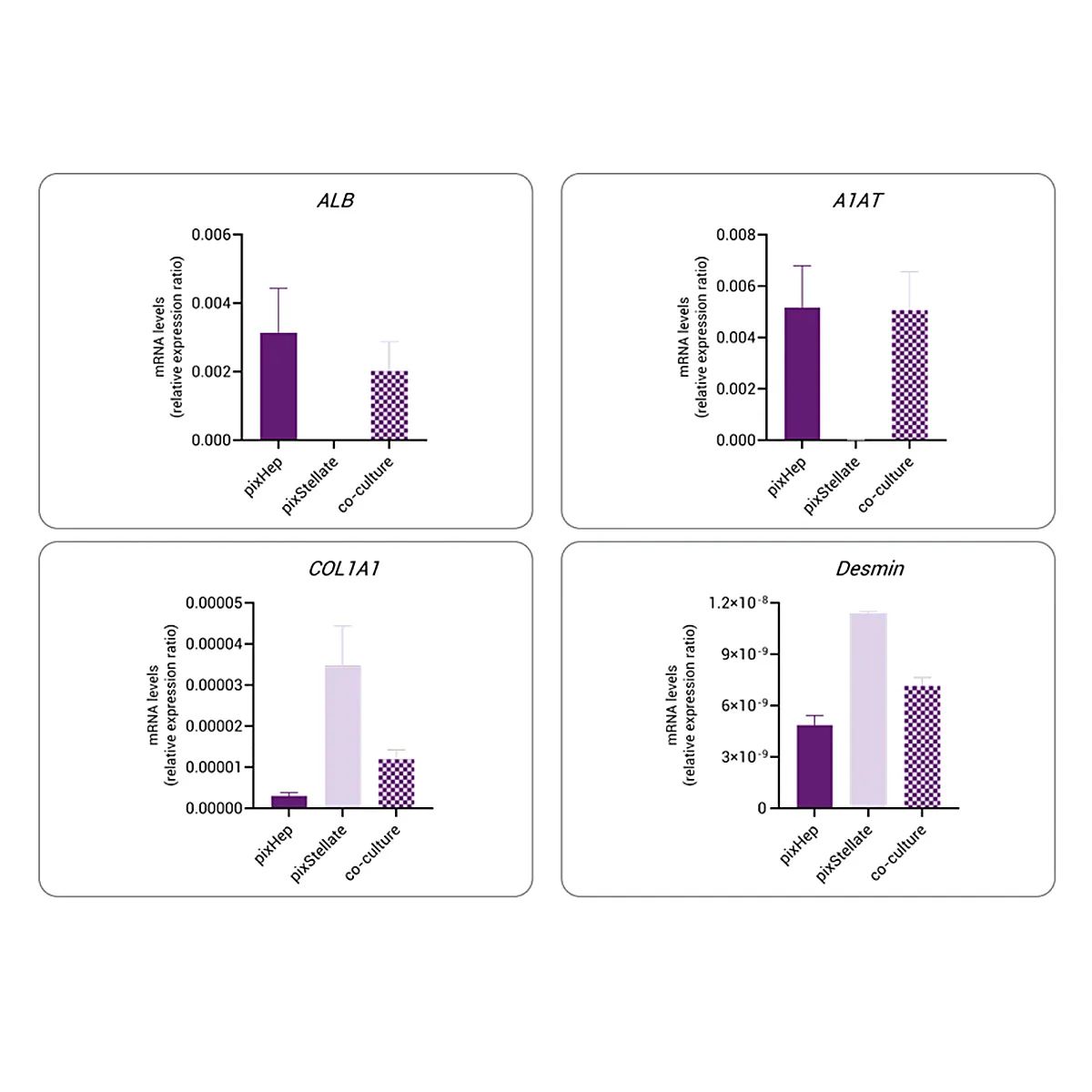
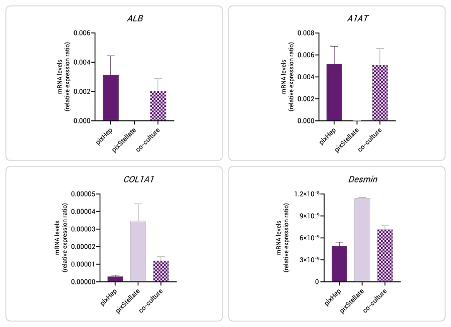
- pixHep (HEP-003, 1 × 10⁶ cells per cryopreserved vial) and pixStellate (HSC-003) optimized for advanced, donor matched co-culture studies (1 cryopreserved vial of each required
Place your Order
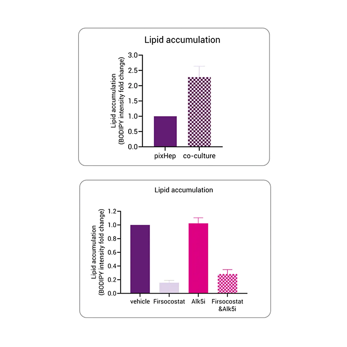
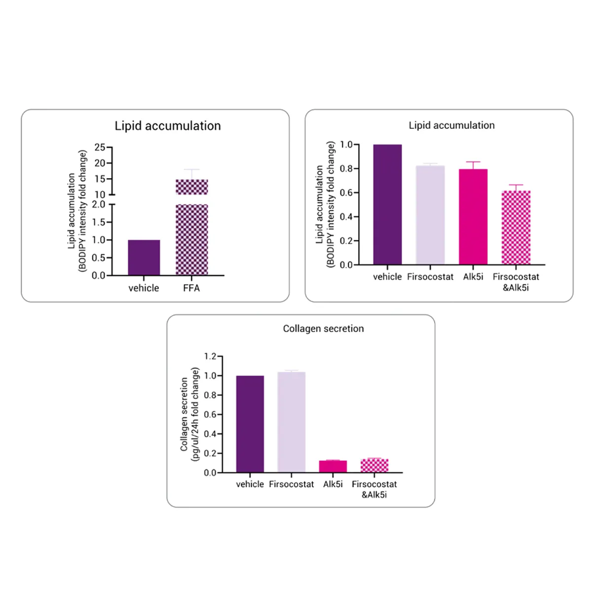
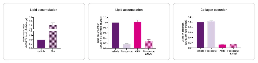
This model recreates the progression from MASLD to fibrotic MASH by combining lipid-loaded pixHep™ with activated pixStellate™ cells.
Under steatogenic and pro-inflammatory conditions, hepatocytes accumulate fat and signal stellates to deposit collagen and upregulate fibrotic genes.
Integrated lipid, inflammatory, and fibrotic readouts.
Quantifiable collagen deposition and α-SMA upregulation.
Compatible with anti-fibrotic, metabolic, and anti-inflammatory drugs.
CRISPR-enabled for genetic risk variant modelling.
Technical Data & Functional Validation
Key HSC and Hepatocyte Markers (mRNA)
Hepatic Steatosis and Pharmacological Reversal

Fatty Acid Treatment and Collagen Secretion

Streamline Your Research
Ready to Turn Your Cells Into Data?
Our pixCell portfolio seamlessly integrates into custom data generation projects—from functional assays (pixCellServices) to phenomic analysis (pixCellPaint)—delivering ready-to-use data at your fingertips.

