


Catalog Number: MAC-003
pixMac iPSC-derived Macrophage Cells
Human iPSC-derived macrophage cells (1 × 10⁶ viable cells per vial) for modeling immunity and inflammation.
- Express key macrophage identity markers (CD14, CD68) and polarization marker MRC1/CD206
- Respond to IFNγ and LPS or IL-4 stimulation to adopt pro-inflammatory (M1) or anti-inflammatory (M2) phenotypes
- Donor-matched co-culture compatible with pixHep for MASLD and steatohepatitis modelling
pixMac™ are iPSC-derived macrophages that express key macrophage identity markers (CD14, CD68) and the polarisation marker MRC1/CD206, and respond to inflammatory or anti-inflammatory cues.
They can be used alone or paired with pixHep™, in donor-matched co-culture, to model immune-driven liver injury and MASLD-to-steatohepatitis progression.
Unlimited supplyDerived from renewable iPSCs, pixMac removes the donor-to-donor variability and finite availability of primary blood-derived macrophages.
Defined & reproducibleA standardised differentiation workflow yields a consistent naive (M0) population, batch after batch.
Fully polarisablepixMac responds to cues to adopt pro-inflammatory (M1) or anti-inflammatory (M2) states, validated at mRNA and protein level.
Application-readySuited to inflammation, infectious disease, complex cell modelling and drug-safety screening.
Technical Data & Functional Validation
pixlbio iPSC-derived macrophages (pixMac) express key monocyte and macrophage identity markers, respond to phagocytic stimuli, and mount phenotype-specific transcriptional and secretory responses on M1/M2 polarisation.
The pixMac differentiation workflow
.png)
Pluripotent stem cells (iPSC) undergo monocytopoiesis for long-term monocyte output. Monocytes are harvested and seeded, then differentiated into a naive (M0) macrophage population, which can be polarised toward a pro-inflammatory (M1, host defence and tumour surveillance) or anti-inflammatory (M2, tissue repair, resolution and immune modulation) phenotype.
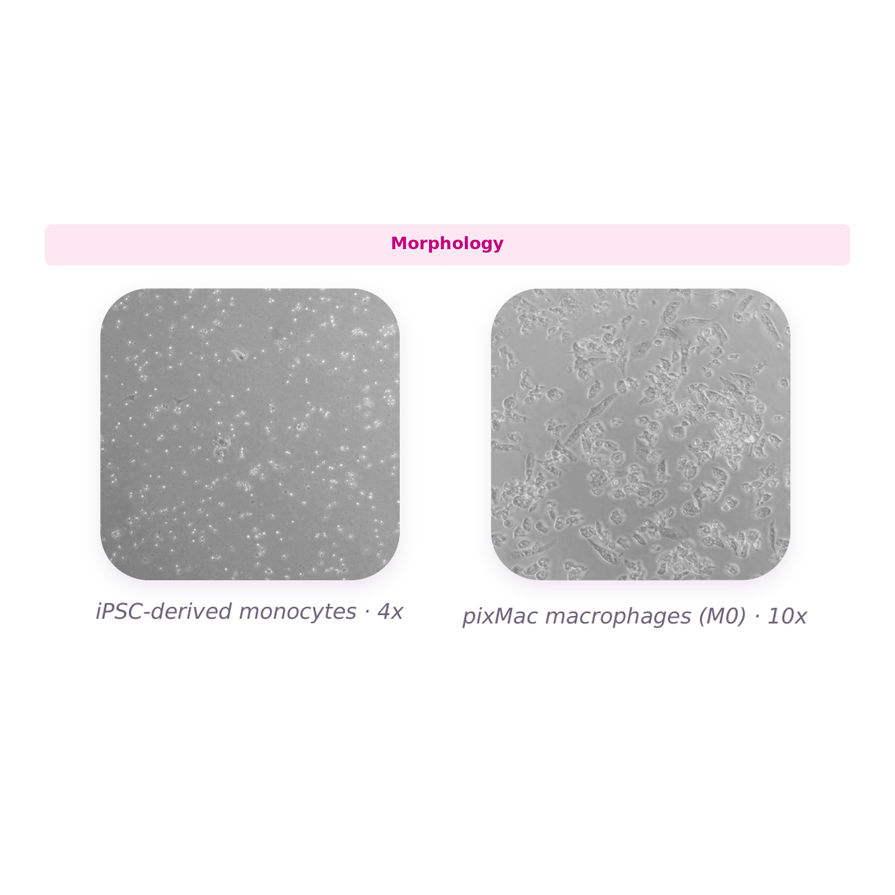
Morphology
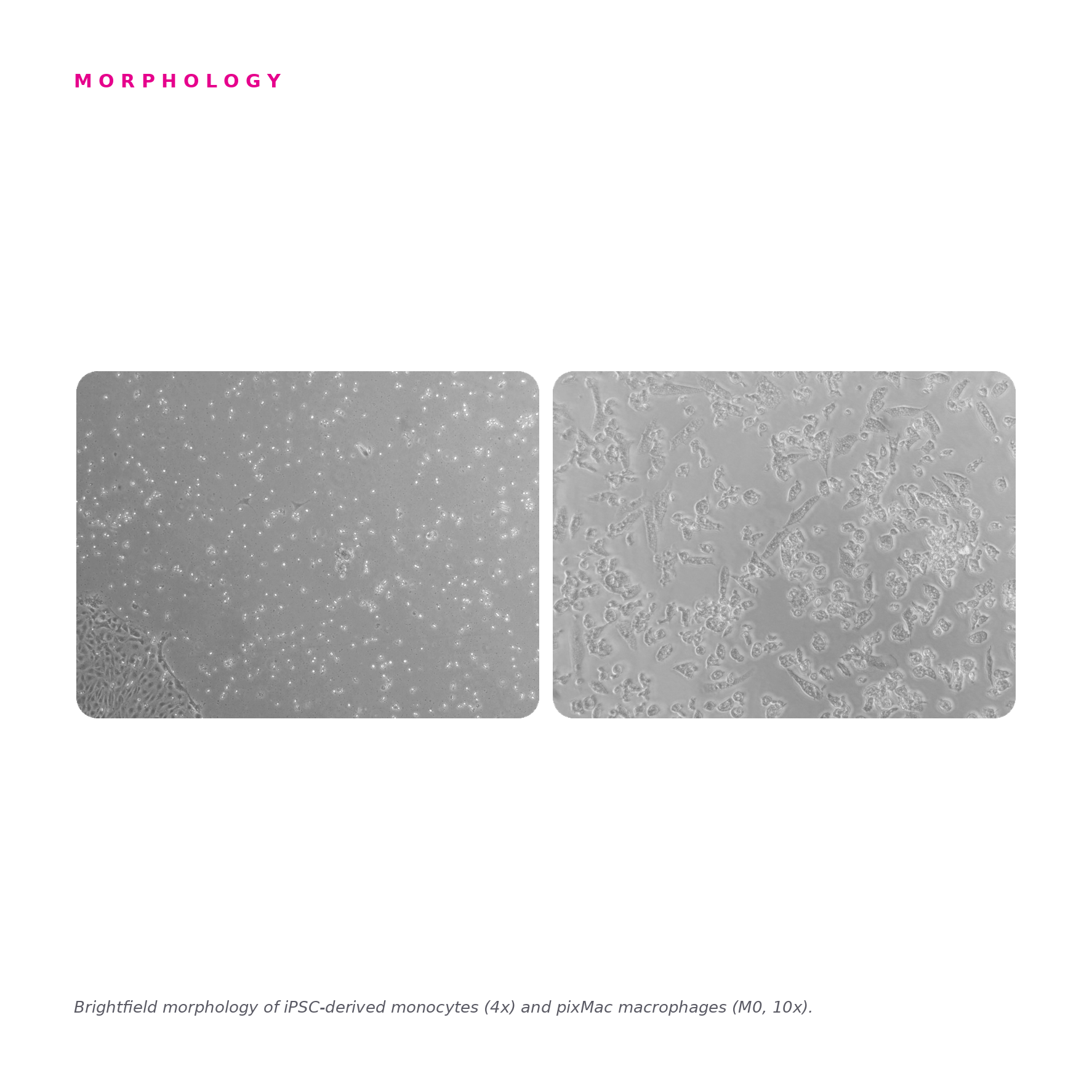
Brightfield morphology of iPSC-derived monocytes (4x) and pixMac macrophages (M0, 10x), showing the characteristic transition from small, rounded monocytes to larger, adherent, vacuolated macrophages.
Marker expression (qPCR)
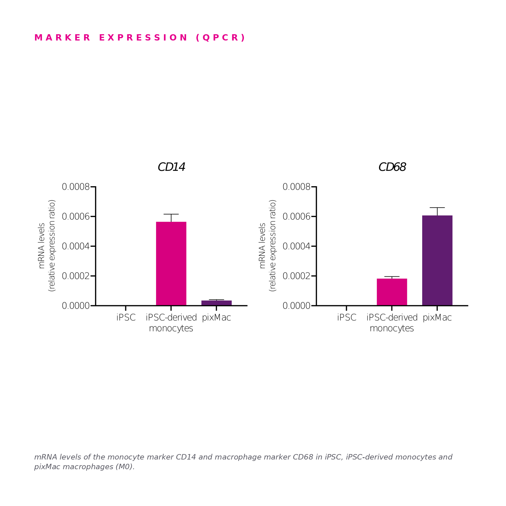
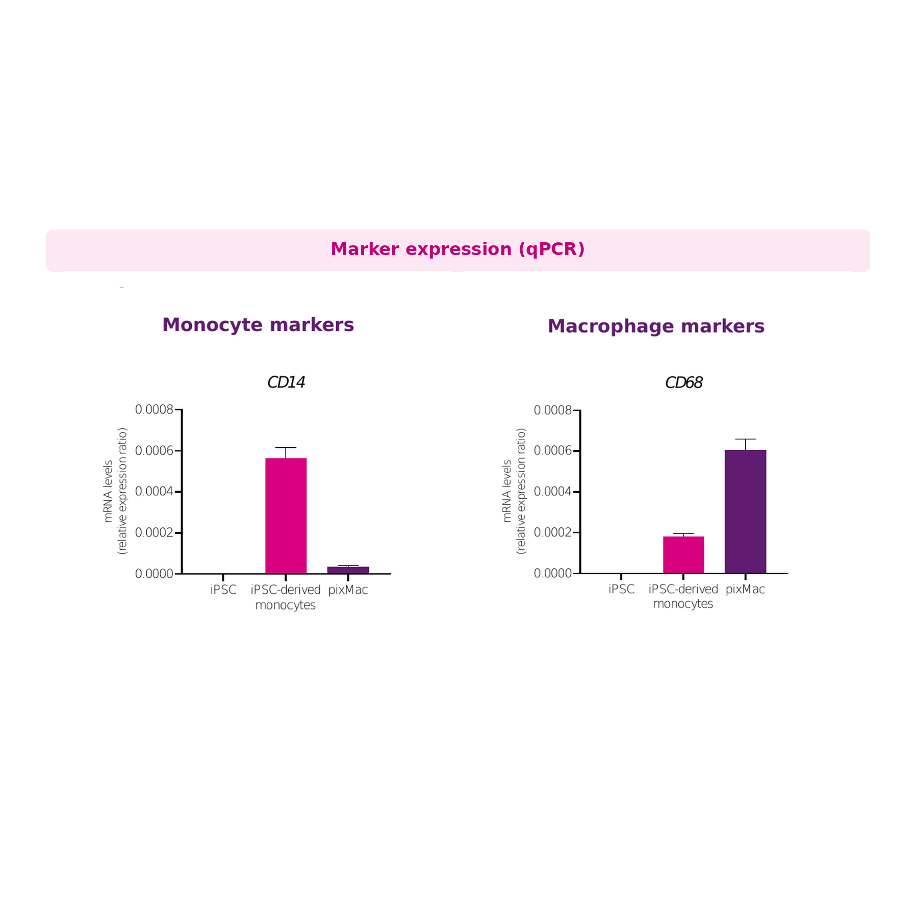
mRNA levels of the monocyte marker CD14 and macrophage marker CD68 in iPSC, iPSC-derived monocytes, and pixMac macrophages (M0). CD14 is enriched in the monocyte stage, while CD68 rises further on macrophage differentiation. Data presented as mean±SD of n=2 biological replicates; mRNA expression normalised to 18S sRNA housekeeping gene.
Immunofluorescence
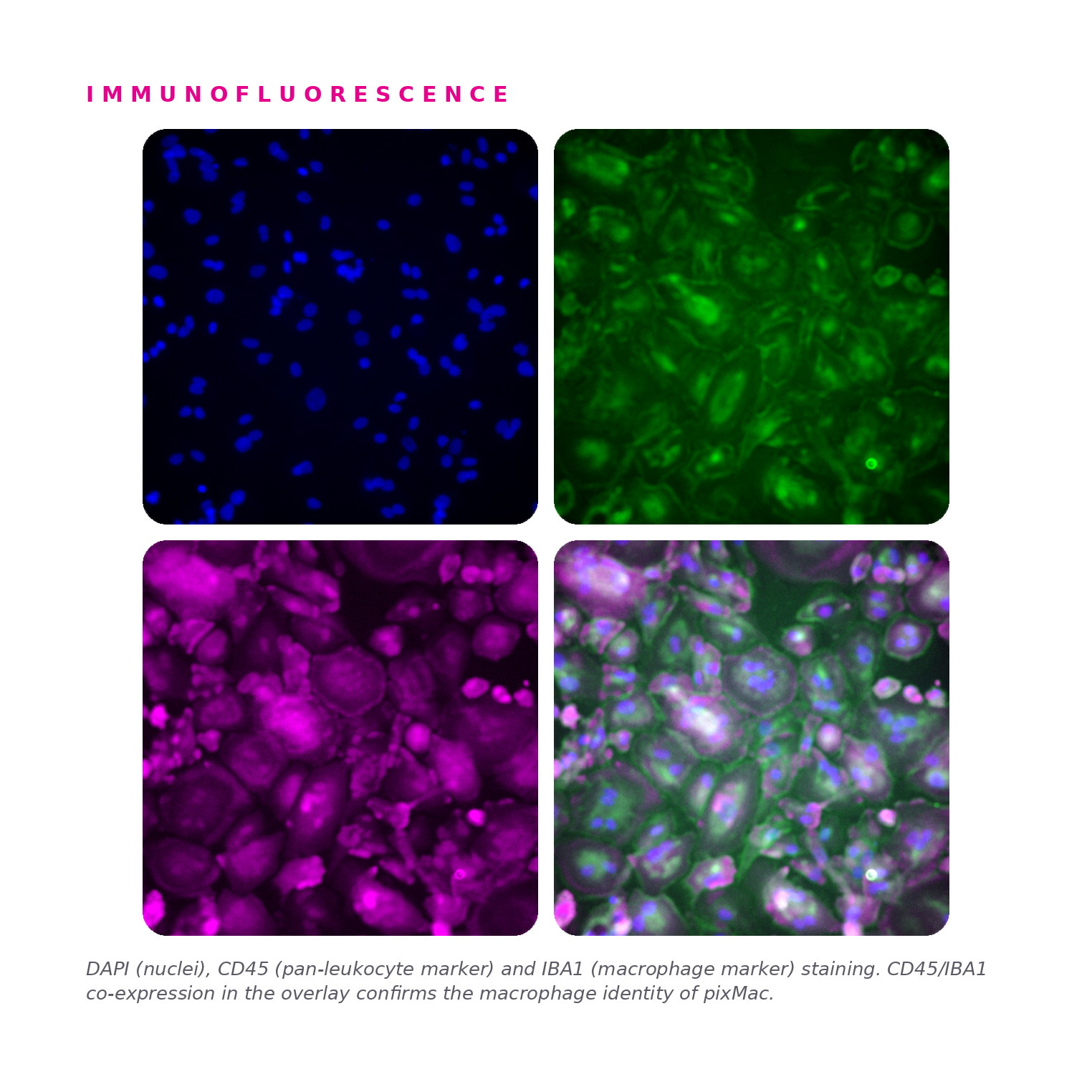
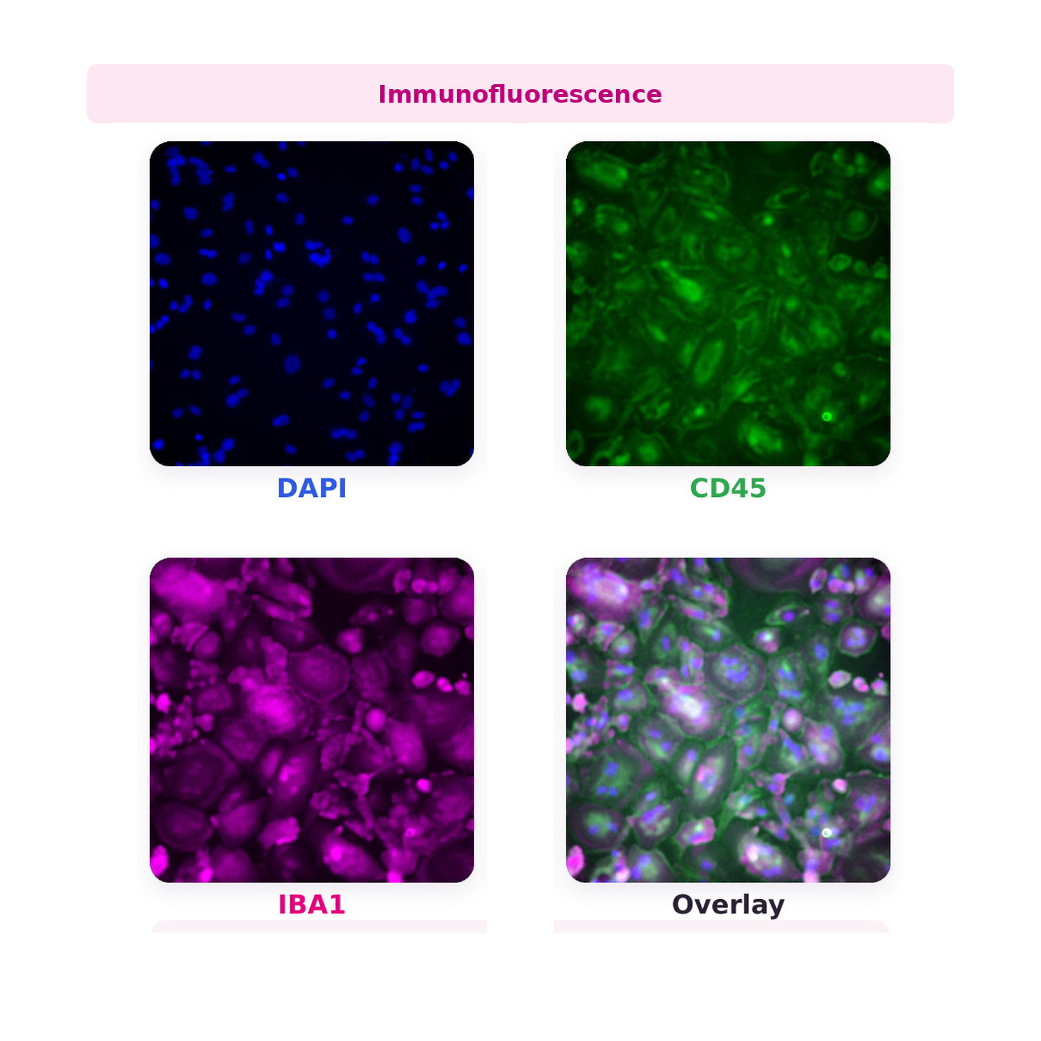
Immunofluorescence staining for DAPI (nuclei), CD45 (pan-leukocyte marker) and IBA1 (macrophage marker) in pixMac. CD45 and IBA1 co-expression in the overlay confirms the macrophage identity of pixMac.
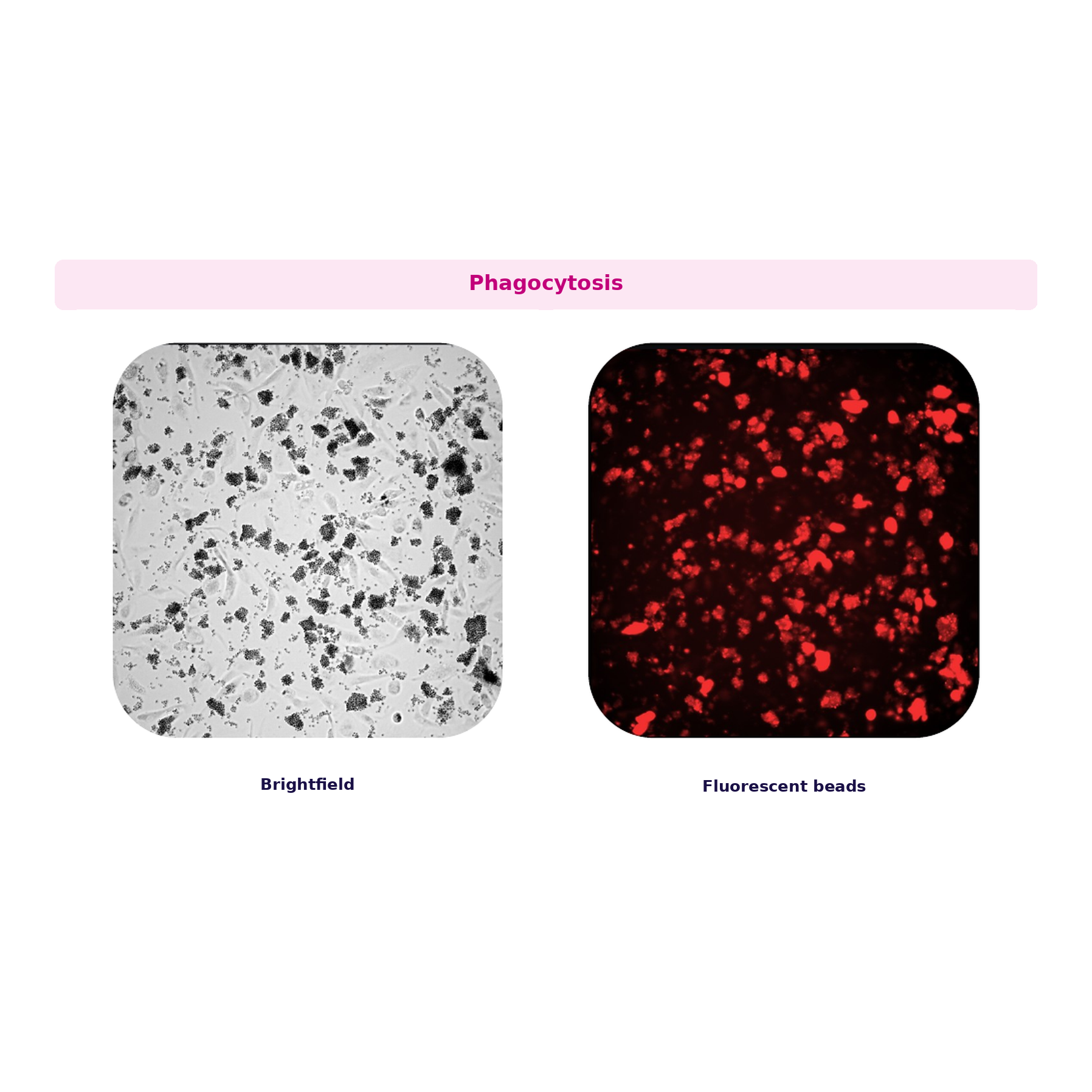
Phagocytosis
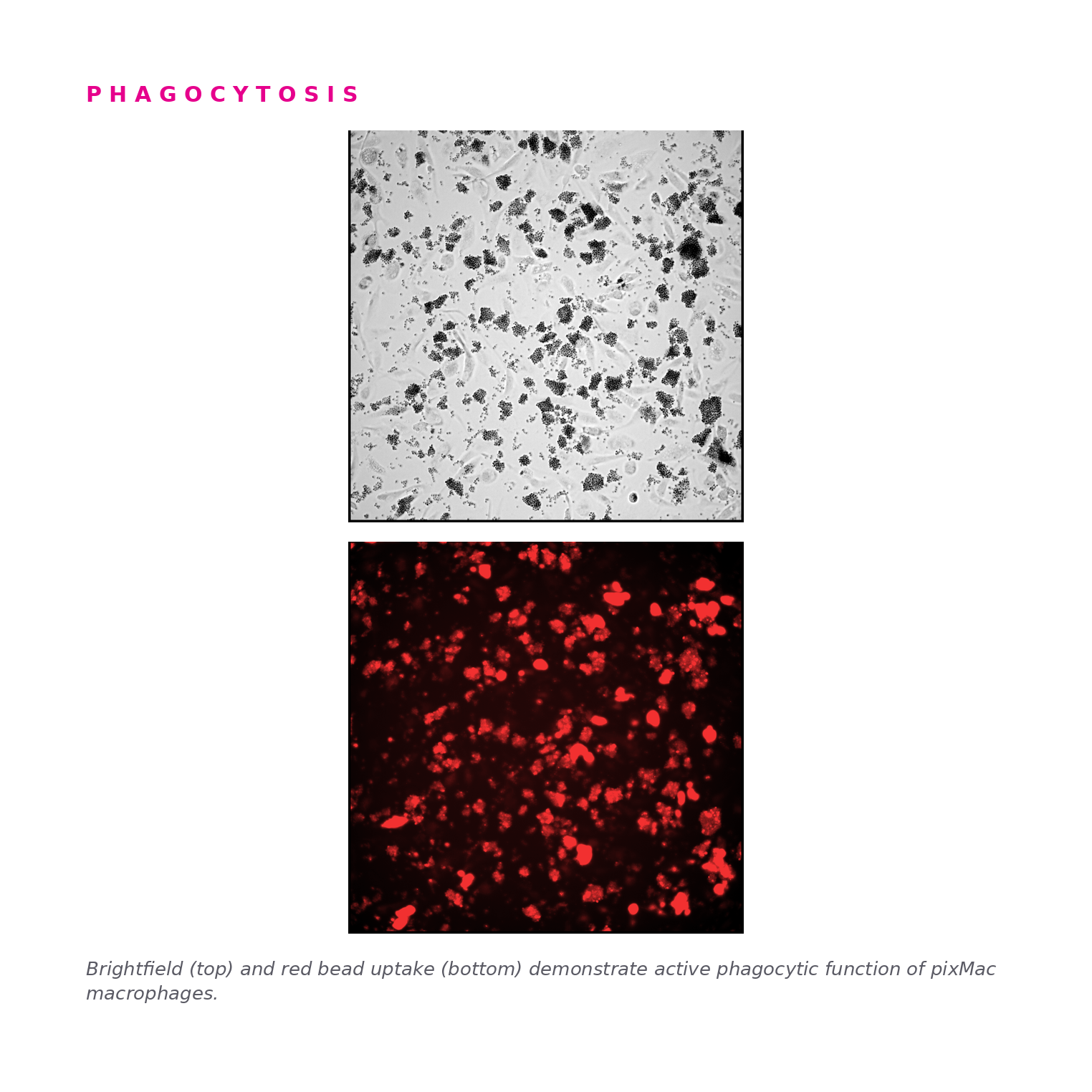
Brightfield (top) and fluorescent red bead uptake (bottom) demonstrating active phagocytic function of pixMac macrophages.
Functional polarisation: M1 (pro-inflammatory) response
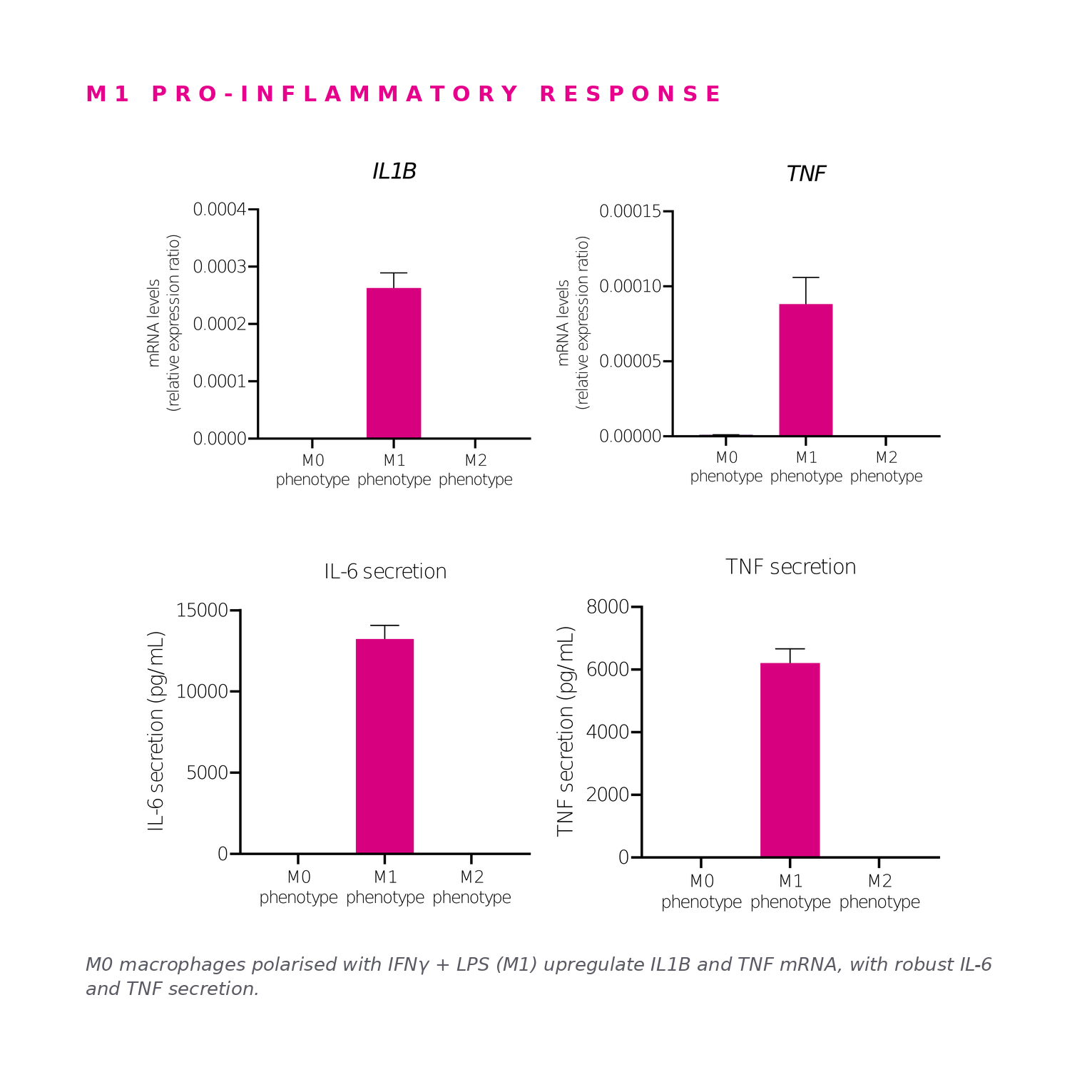
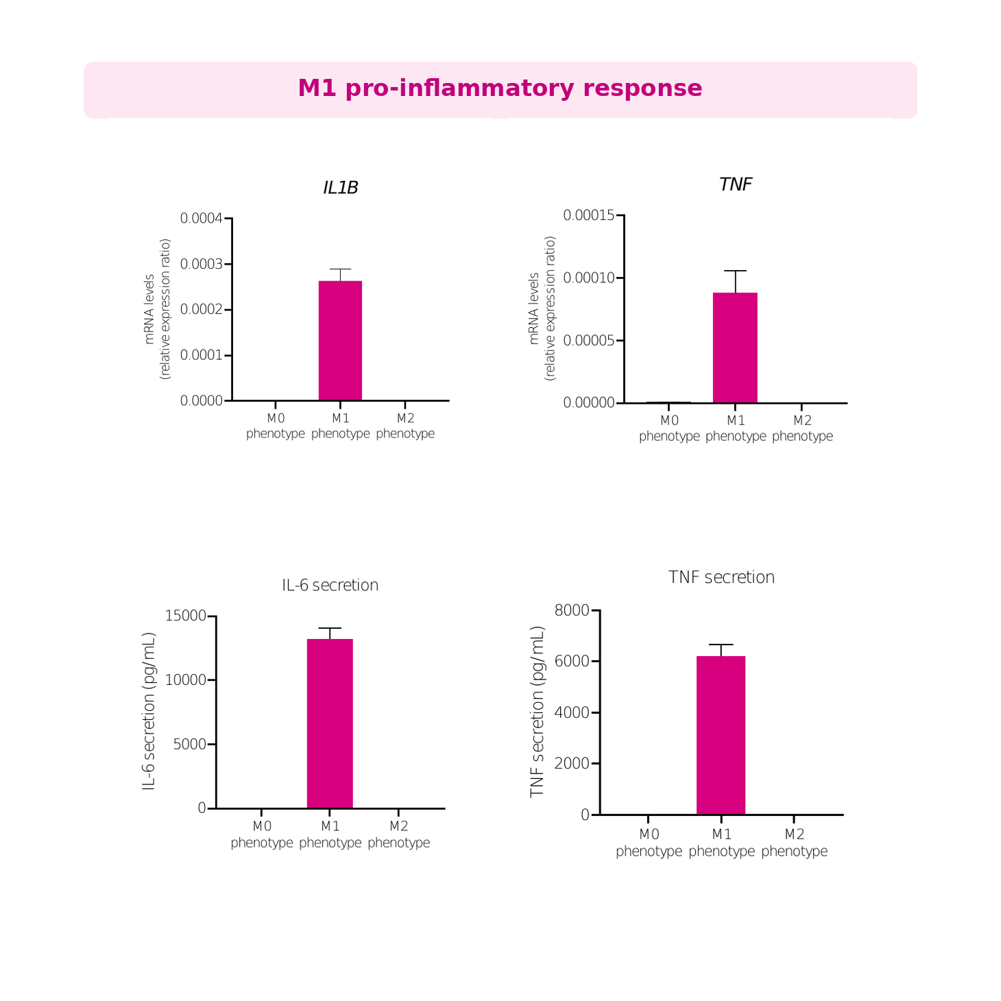
M0 macrophages polarised for 24 h with IFNγ + LPS (M1) upregulate IL1B and TNF mRNA, with robust IL-6 (~13,000 pg/mL) and TNF (~6,000 pg/mL) secretion. Data presented as mean±SD of n=2 biological replicates.
Functional polarisation: M2 (anti-inflammatory) response
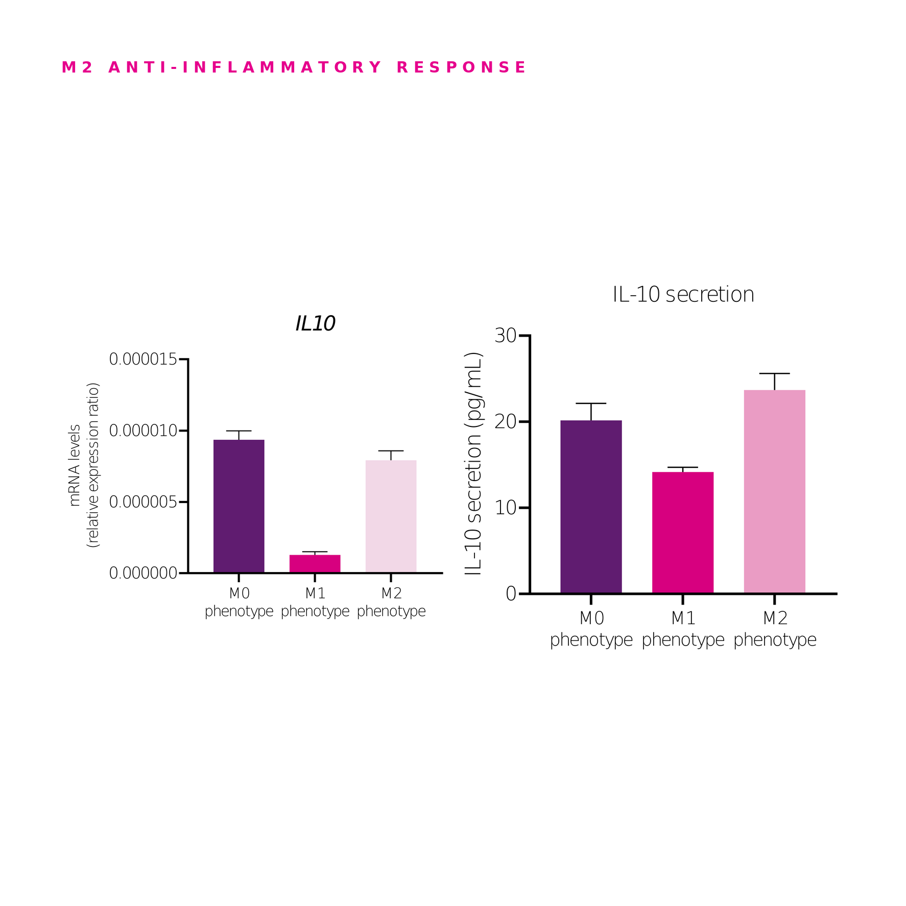
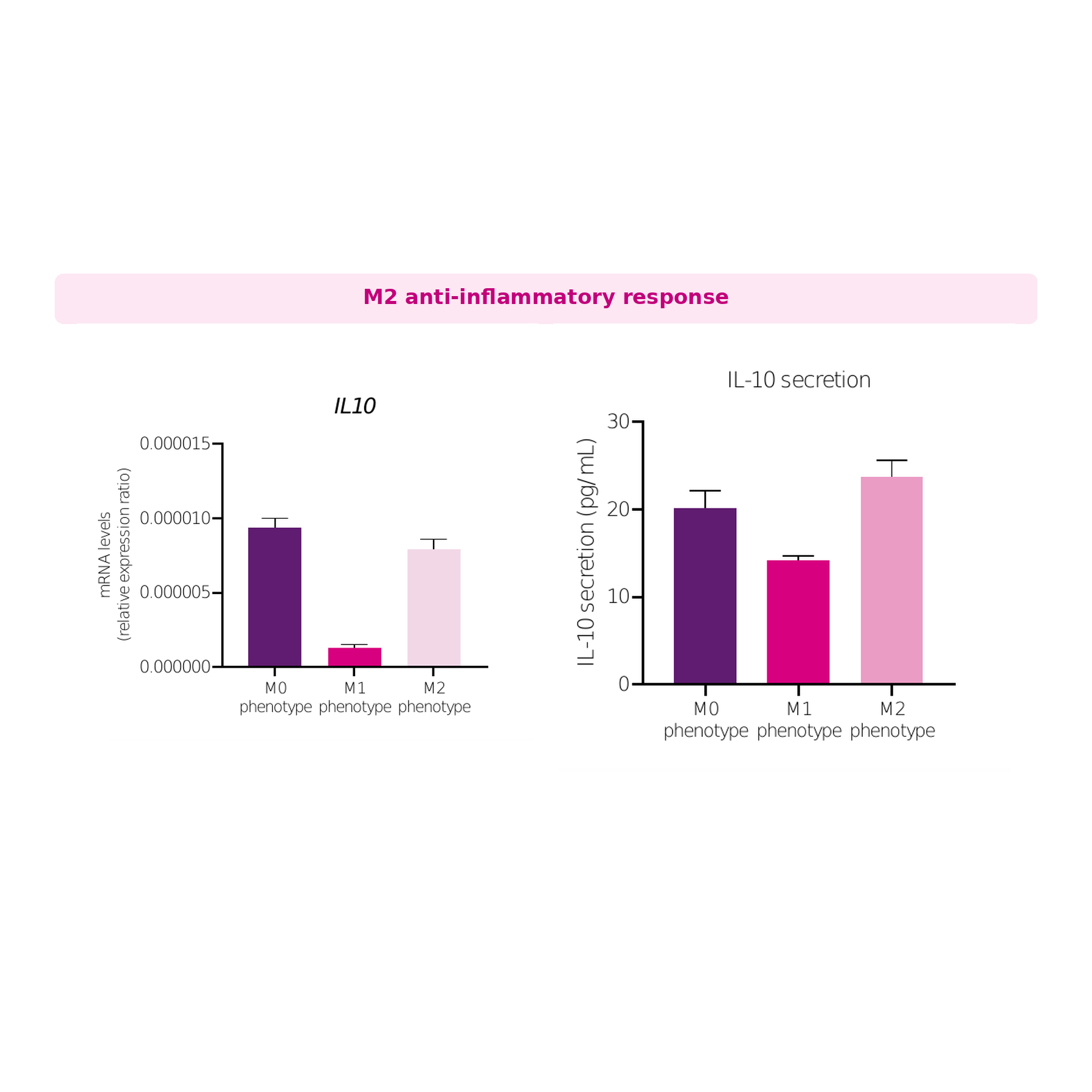
M0 macrophages polarised for 24 h with IL-4 (M2) restore IL10 mRNA and secreted protein (~24 pg/mL secreted) relative to M1, which suppresses IL10 at both mRNA and protein level. Phenotype-specific responses confirm pixMac as a tunable model of macrophage polarisation. Data presented as mean±SD of n=2 biological replicates.
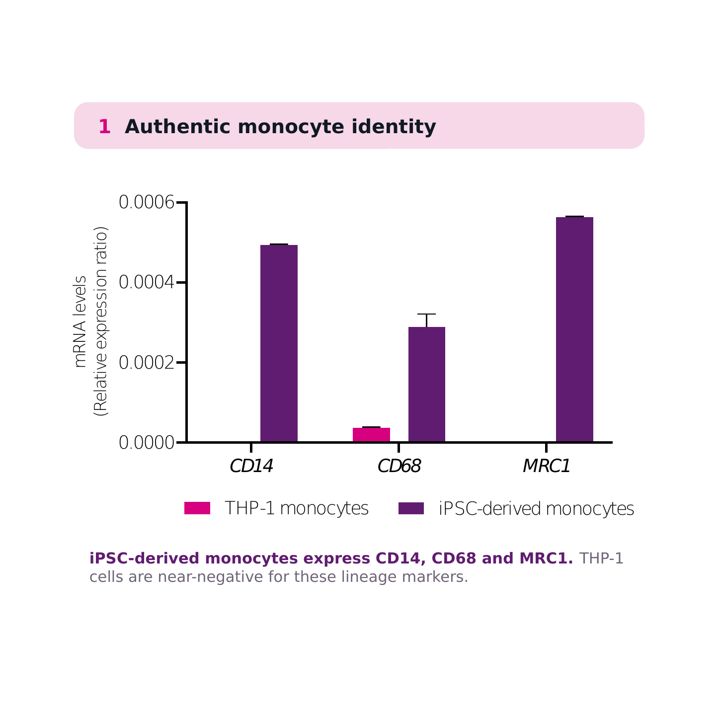
Beyond immortalised cell lines: authentic monocyte identity
.png)
iPSC-derived monocytes express CD14, CD68 and MRC1. THP-1 monocytic cells are near-negative for these lineage markers. Data presented as mean±SD of n=2 biological replicates.
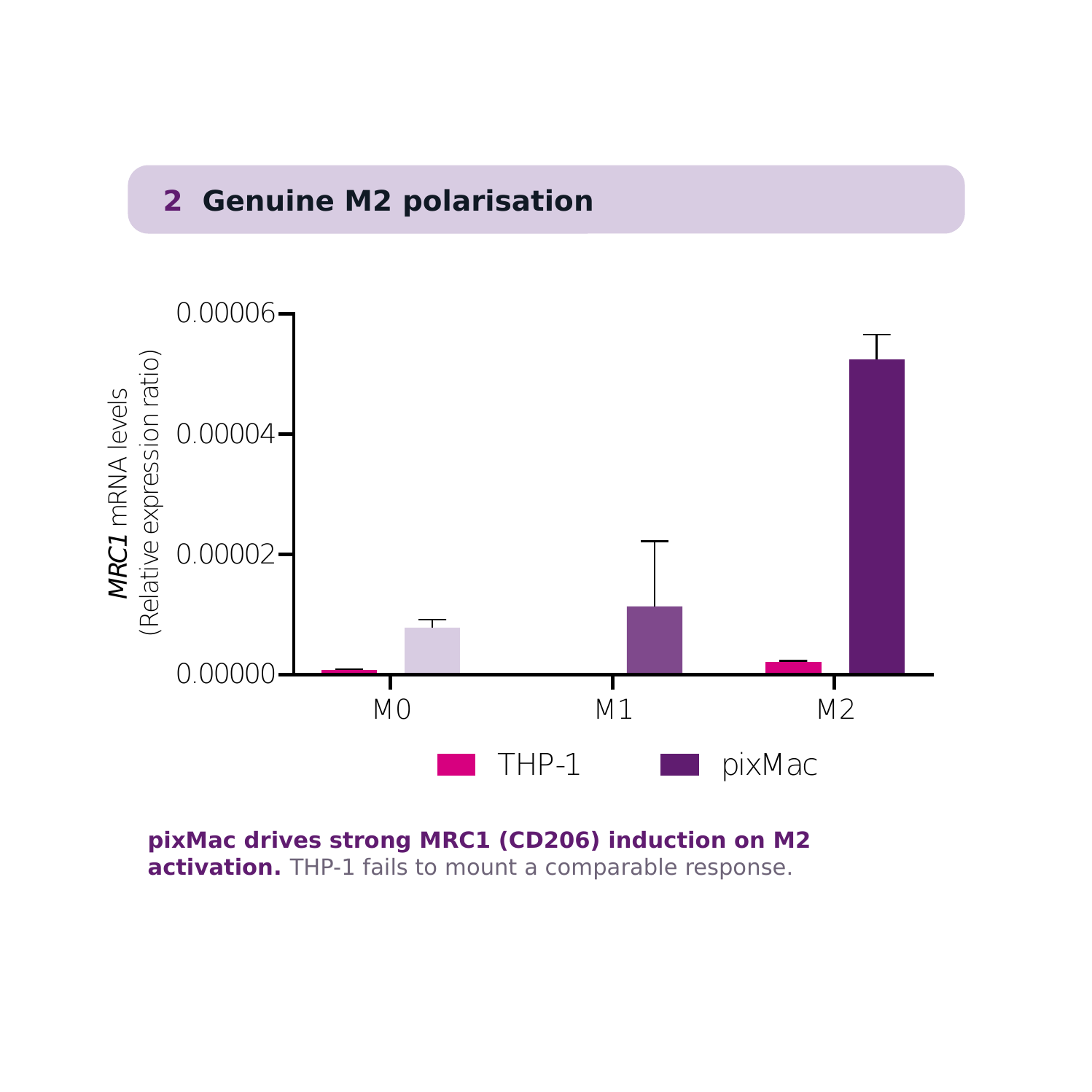
Beyond immortalised cell lines: genuine M2 polarisation
.png)
pixMac drives strong MRC1 (CD206) induction on M2 activation. THP-1 fails to mount a comparable response, underscoring pixMac's advantage over immortalised monocytic lines for polarisation studies. Data presented as mean±SD of n=2 biological replicates.
Ready to Turn Your Cells Into Data?